Frisen scale grading describes the severity of papilledema in six stages, numbered 0 to 5.
Lars Frisén published this staging scheme in 1982, and clinicians still use it at the slit lamp.
However, the grade describes disc appearance only, so it cannot establish the cause or the visual prognosis.
What the Frisen Scale Measures
The scale records visible changes at the optic nerve head and peripapillary nerve fiber layer.
Raised intracranial pressure impairs axoplasmic flow, and the axons then swell.
As a result, the examiner sees a gray halo, blurred margins and obscured vessels.
Before You Assign a Grade
First, confirm that the swelling is true papilledema (see papilledema basics).
Next, examine the dilated disc with a stereoscopic view, such as a 78D or 90D lens.
Also, grade each eye separately, since asymmetry is common.
Fundus Explorer Pro
Photograph the retinal findings described here with the phone already in your pocket — 22 D optics and built-in illumination in one handheld unit.
From Choroida — the team behind this siteStage 0: Normal Optic Disc
The nerve fiber layer keeps its radial pattern, and its striations show no tortuosity.
The disc borders may look less sharp nasally, superiorly and inferiorly.
Therefore, a blurred upper margin alone should not prompt a higher grade.
Stage 1: Very Early Papilledema
A subtle grayish C-shaped halo surrounds the disc, with a gap on the temporal side.
This halo disrupts the normal radial nerve fiber striations.
Red-free light can help, because it makes these faint changes easier to see.
Stage 2: Early Papilledema
The halo now becomes circumferential and closes the earlier temporal gap.
The nasal border also becomes elevated, while the major vessels remain visible.
For example, transient visual obscurations can occur while central vision tests normally.
Stage 3: Moderate Papilledema
Vessel obscuration defines this stage, so inspect the major vessels as they leave the disc.
One or more segments disappear beneath the swollen nerve fibers at the disc border.
In addition, elevation now involves all borders of the disc.
The halo often develops an irregular outer fringe with finger-like extensions.
Stage 4: Marked Papilledema
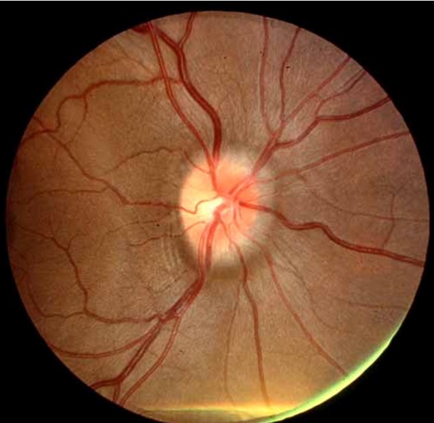
Here, swelling elevates the entire nerve head and obscures its borders.
A segment of a major vessel also disappears on the disc surface itself.
This location separates stage 4 from stage 3, so trace each vessel carefully.
Stage 5: Severe Papilledema
The nerve head expands forward in a dome-shaped protrusion and obliterates the physiologic cup.
Then the peripapillary halo becomes narrow and smoothly demarcated.
Consequently, the dome shape carries more weight than vessel findings at this stage.
Signs Outside the Frisen Scale
Hemorrhages, cotton wool spots and exudates do not define any stage.
Paton lines, the circumferential peripapillary folds, also fall outside the criteria.
Nevertheless, these findings support true disc swelling, so record them separately.
Limits of the Frisen Scale
The stages form an ordinal scale, so the step between grades is not a fixed amount of swelling.
Also, the grade does not measure intracranial pressure directly.
A low grade cannot exclude raised pressure, because some patients have little or no swelling.
Finally, optic atrophy can reduce visible swelling while function continues to decline.
Therefore, check acuity, color vision, pupils and perimetry at each visit, as the IIH Treatment Trial used perimetric mean deviation as its primary outcome.
Frisen Scale Reliability
Grading remains subjective, and published evaluations report imperfect agreement between raters.
Adjacent stages cause the most trouble because their boundaries can look similar.
Therefore, side-by-side photographs give a fairer comparison than memory or an old chart note.
Discs That Confuse Grading
Crowded, tilted and myopic discs can mimic blurred margins.
Drusen also elevate the disc, although the vessels usually remain clearly visible over it (see pseudopapilledema).
Moreover, inflammatory and ischemic disc swelling can share similar signs (see other causes of disc swelling).
The scale was built for staging swelling, so it cannot separate these diagnoses.
OCT Alongside the Frisen Scale
OCT adds a continuous measure of peripapillary and retinal thickness.
Scott and colleagues found that OCT thickness measures related to clinical grading in raised intracranial pressure.
However, segmentation can fail in severe swelling, so review each scan for errors.
Thinning may also reflect atrophy, and ganglion cell analysis can help clarify this.
Explaining Frisen Scale Grades to Patients
Patients often assume that a grade directly predicts their chance of vision loss.
Instead, explain that it describes how swollen the nerve looks today.
Also, advise urgent review for new visual loss, because symptoms may change before the next appointment.
Pitfalls to Avoid
- Do not grade a swollen disc before considering pseudopapilledema
- Avoid confusing vessel obscuration at the border with obscuration on the disc
- Never treat a lower grade as proof of recovery without visual field testing
- Do not use a low grade to exclude raised intracranial pressure


Document what you see
Two smartphone imaging tools built for everyday clinic use — one for the slit lamp, one for the fundus.
From Choroida — the team behind this siteReferences
- Frisén L. Swelling of the optic nerve head: a staging scheme. J Neurol Neurosurg Psychiatry. 1982;45:13-18.
- Scott CJ, Kardon RH, Lee AG, Frisén L, Wall M. Diagnosis and grading of papilledema in patients with raised intracranial pressure using optical coherence tomography vs clinical expert assessment using a clinical staging scale. Arch Ophthalmol. 2010;128:705-711.
- Sinclair AJ, Burdon MA, Nightingale PG, et al. Rating papilloedema: an evaluation of the Frisén classification in idiopathic intracranial hypertension. J Neurol. 2012;259:1406-1412.
- Wall M, McDermott MP, Kieburtz KD, et al. Effect of acetazolamide on visual function in patients with idiopathic intracranial hypertension and mild visual loss. JAMA. 2014;311:1641-1651.

Writes and reviews the clinical articles on Educate. Every post is checked against peer-reviewed sources before it goes live.