SUN classification is the Standardization of Uveitis Nomenclature, a shared system for naming and grading intraocular inflammation.
It defines anatomic type, onset, duration and course, and it sets grading scales for cells and flare.
Therefore, it lets clinicians and researchers describe the same eye in the same words.
Why the SUN Classification Exists
Before 2005, uveitis terms varied between centers and countries.
Comparing outcomes was difficult, and trial data were hard to pool.
Also, the Standardization of Uveitis Nomenclature working group set definitions at a workshop.
Because a common language helps care, the system became widely used.
Anatomic Types Under the SUN Classification
Anterior uveitis involves the anterior chamber primarily.
Intermediate uveitis involves the vitreous, and posterior uveitis involves the retina or choroid.
Also, panuveitis involves all three compartments.
Therefore, the first step is to name the primary site of inflammation.
All-fit Slit-Lamp Adapter
Record and share exactly what you see at the slit lamp. One adapter fits any slit lamp or surgical microscope — and any smartphone.
From Choroida — the team behind this siteSubtypes of Anterior Uveitis
Iritis describes inflammation of the iris alone.
Iridocyclitis involves the iris and the pars plicata.
Also, anterior cyclitis involves the ciliary body mainly (see acute anterior uveitis).
Because the terms reflect the focus, use the one that fits.
Intermediate and Posterior Types
Pars planitis is a subtype of intermediate uveitis with snowbanking (see pars planitis).
Posterior uveitis includes focal, multifocal and diffuse choroiditis, retinitis and retinal vasculitis.
Also, panuveitis shows disease in the anterior chamber, the vitreous and the retina or choroid.
Therefore, anatomy narrows the differential.
Onset in the SUN Classification
Onset can be sudden or insidious.
Sudden onset suggests an acute anterior uveitis such as HLA-B27 disease.
Also, insidious onset suggests chronic disease such as juvenile idiopathic arthritis uveitis.
Because the pattern aids diagnosis, record it.
Duration and Course in the SUN Classification
Limited disease lasts up to three months, and persistent disease lasts longer.
Acute describes a sudden onset with limited duration.
Recurrent disease has repeated episodes separated by at least three months without treatment.
Also, chronic disease relapses within three months of stopping treatment.
Grading Anterior Chamber Cells
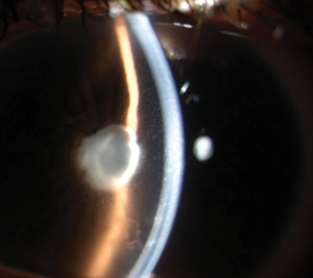
Count cells in a one-millimeter by one-millimeter beam.
Grade zero has fewer than one cell, and 0.5 plus has one to five cells.
Also, one plus has six to fifteen, two plus has sixteen to twenty-five, three plus has twenty-six to fifty and four plus has more than fifty.
Therefore, a standard method allows comparison between visits.
Grading Flare
Flare is graded from zero to four plus.
Zero means none and one plus means faint.
Also, two plus is moderate with clear iris and lens details, three plus is marked with hazy details and four plus is intense with fibrin.
Because flare reflects protein, it is less reliable than cells for activity.
Grading Vitreous Haze
Vitreous haze is graded by the clarity of fundus details.
The National Eye Institute scale runs from zero to four plus.
Also, observers differ in grading, so use photographs as a reference.
Therefore, apply the same method at each visit.
Defining Activity in the SUN Classification
Inactive disease means zero cells.
Worsening activity means a two-step increase in cells or haze, or an increase to three plus.
Also, improved activity means a two-step decrease.
Remission means inactive disease for at least three months off treatment.
Using the SUN Classification in Practice
Record the type, onset, duration and course in each note.
Also, grade cells and flare at each visit.
Because clear notes support decisions, use the system routinely.
Limits
The classification is descriptive and does not name the cause.
Also, grading depends on the examiner and the light.
Therefore, combine the system with the etiologic work-up.
Use in Research
Trials use the system to define inclusion and endpoints.
A standard grading scale allows pooling of data.
Also, registries use the terms to compare outcomes.
Because the language is shared, the evidence is easier to interpret.
Examples of Classification
A young man with a sudden painful red eye and cells in the anterior chamber has acute anterior uveitis.
A woman with a persistent vitritis and snowbanking has intermediate uveitis.
Also, a patient with retinal infiltrates and vitreous cells has a posterior or panuveitis.
Therefore, the system turns the examination into a short label.
Limitations at the Slit Lamp
Cell grading varies with the beam size and the room light.
Also, low-grade inflammation may be hard to grade.
Because the examiner affects the result, use the same method at each visit.
For the latest studies, see the PubMed results for SUN classification.


Document what you see
Two smartphone imaging tools built for everyday clinic use — one for the slit lamp, one for the fundus.
From Choroida — the team behind this siteReferences
- Jabs DA, Nussenblatt RB, Rosenbaum JT; Standardization of Uveitis Nomenclature (SUN) Working Group. Standardization of uveitis nomenclature for reporting clinical data: results of the First International Workshop. Am J Ophthalmol. 2005;140:509-516.
- Trusko B, Thorne J, Jabs D, et al. The Standardization of Uveitis Nomenclature (SUN) Project: development of a clinical evidence base utilizing informatics tools and techniques. Methods Inf Med. 2013;52:259-265.
- Bloch-Michel E, Nussenblatt RB. International Uveitis Study Group recommendations for the evaluation of intraocular inflammatory disease. Am J Ophthalmol. 1987;103:234-235.
- Deschenes J, Murray PI, Rao NA, Nussenblatt RB. International Uveitis Study Group (IUSG): clinical classification of uveitis. Ocul Immunol Inflamm. 2008;16:1-2.

Writes and reviews the clinical articles on Educate. Every post is checked against peer-reviewed sources before it goes live.