
CASE REPORT
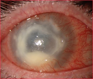
Case of Acanthamoeba Keratitis a 38-year-old female was admitted to the ophthalmology department in a hospital with pain, redness, watering, and irritation of the left eye. She gave a history of paddy husk injury to the left eye 7 days prior to the development of these symptoms and also noticed a white spot in the left eye 4 days after the injury.
On slit-lamp examination, the left eye showed circumcorneal congestion with a central corneal ulcer of 1.6 x 1.4 mm size with clear margins extending into the superficial stroma. The anterior chamber was normal.
There was no hypopyon. Lacrimal passages were free. Visual acuity in the right eye was 6/6 whereas in the left eye it was 6/18. Multiple corneal ulcer scrapings stained with Giemsa stain showed cysts of Acanthamoeba which was confirmed by culture. No bacteria or fungi were isolated from these corneal ulcer scrapings.
DISEASE of Acanthamoeba Keratitis
Acanthamoeba keratitis, first recognized in 1973, is a rare, vision-threatening, parasitic infection seen most often in contact lens wearers.
It is often characterized by pain out of proportion to findings and the late clinical appearance of a stromal ring-shaped infiltrate. It is both difficult to diagnose and difficult to treat.
All-fit Slit-Lamp Adapter
Record and share exactly what you see at the slit lamp. One adapter fits any slit lamp or surgical microscope — and any smartphone.
From Choroida — the team behind this siteMANAGEMENT of Acanthamoeba Keratitis
Medical treatment for Acanthamoeba keratitis is still evolving. Success has been reported with various combinations of antibiotic, antiviral, antifungal, and antiparasitic drugs.
Different regimens include combinations of diamidines, biguanides, antibiotics, and antifungals. Some topical preparations of diamidines are propamidine-isethionate, hexamidine-di isethionate, and dibromopropamidine.
Biguanides include polyhexamethylene biguanide (PHMB), chlorhexidine. Neomycin-polymyxin B-gramicidin is thought to kill bacteria which provides a food source for the acanthamoeba.
Antifungals include topical and oral preparations of voriconazole as well as ketoconazole, miconazole and clotrimazole .
Read more: Mucormycosis, Plateau Iris, Arcus Senilis, Christmas tree cataract, Crocodile Shagreen


Document what you see
Two smartphone imaging tools built for everyday clinic use — one for the slit lamp, one for the fundus.
From Choroida — the team behind this siteREFERENCES
- Krachmer J, Mannis M, Holland E: CORNEA, 2nd ed.Elsevier Mosby, 2005, 1115-1122.
- Hargrave SL et al: Results of a trial of combined propamidine isethionate and neomycin therapy for Acanthamoeba keratitis. Brolene Study Group, Ophthalmology 106(5):952-957, 1999.
- Bang S, Edell E, Eghrari A, Gottsch J: Treatment with Voriconazole in 3 Eyes with Resistant Acanthamoeba Keratitis. Am Journ Ophthalmol. 2010;149(1):66-69.
- Sun X et al: Acanthamoeba Keratitis: Clinical Characteristics and Management. Ophthalmology 113(3);412-416, 2006.
- Parmar D, Awwad S, Petroll Mm Bowman W, McCulley J, Cavanagh D: Tandem Scanning Confocal Corneal Microscopy in the Diagnosis of Suspected Acanthamoeba Keratitis. Ophthalmology 113(4); 538-547, 2006
- Ocular Pathology Atlas. American Academy of Ophthalmology Web site.
Test yourself
A few questions straight from this article.
-
Which stain of corneal scrapings revealed the cysts that confirmed Acanthamoeba keratitis in this case?
Multiple corneal ulcer scrapings stained with Giemsa showed Acanthamoeba cysts, later confirmed on culture, with no bacteria or fungi isolated. -
Which symptom is characteristic of Acanthamoeba keratitis?
Acanthamoeba keratitis is typically characterised by pain out of proportion to the examination findings, which is one reason it is difficult to diagnose. -
Which corneal sign appears late in the clinical course of Acanthamoeba keratitis?
The late clinical appearance of a stromal ring-shaped infiltrate is a described feature of Acanthamoeba keratitis. -
In which group is Acanthamoeba keratitis seen most often?
Acanthamoeba keratitis is a rare, vision-threatening parasitic infection seen most often in contact lens wearers, though this case followed paddy husk trauma. -
In which year was Acanthamoeba keratitis first recognized?
The article states that Acanthamoeba keratitis was first recognized in 1973. -
What did slit-lamp examination show in this case of Acanthamoeba keratitis?
The left eye showed circumcorneal congestion with a 1.6 x 1.4 mm central corneal ulcer with clear margins extending into the superficial stroma; the anterior chamber was normal with no hypopyon. -
Polyhexamethylene biguanide and chlorhexidine used in Acanthamoeba keratitis belong to which drug class?
PHMB and chlorhexidine are the biguanides used in Acanthamoeba keratitis regimens, alongside diamidines, antibiotics and antifungals. -
Which topical agent used in Acanthamoeba keratitis is a diamidine?
Topical diamidine preparations include propamidine isethionate, hexamidine-diisethionate and dibromopropamidine. -
Why is neomycin-polymyxin B-gramicidin included in regimens for Acanthamoeba keratitis?
Neomycin-polymyxin B-gramicidin is thought to work by killing the bacteria that provide a food source for Acanthamoeba. -
What was the visual acuity of the affected eye in this case of Acanthamoeba keratitis?
Visual acuity was 6/6 in the unaffected right eye and 6/18 in the left eye, which carried the central corneal ulcer.

Writes and reviews the clinical articles on Educate. Every post is checked against peer-reviewed sources before it goes live.


