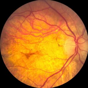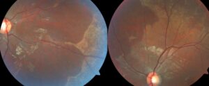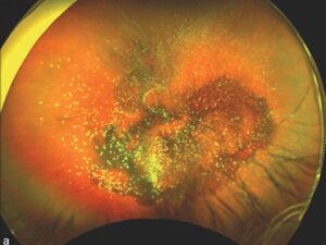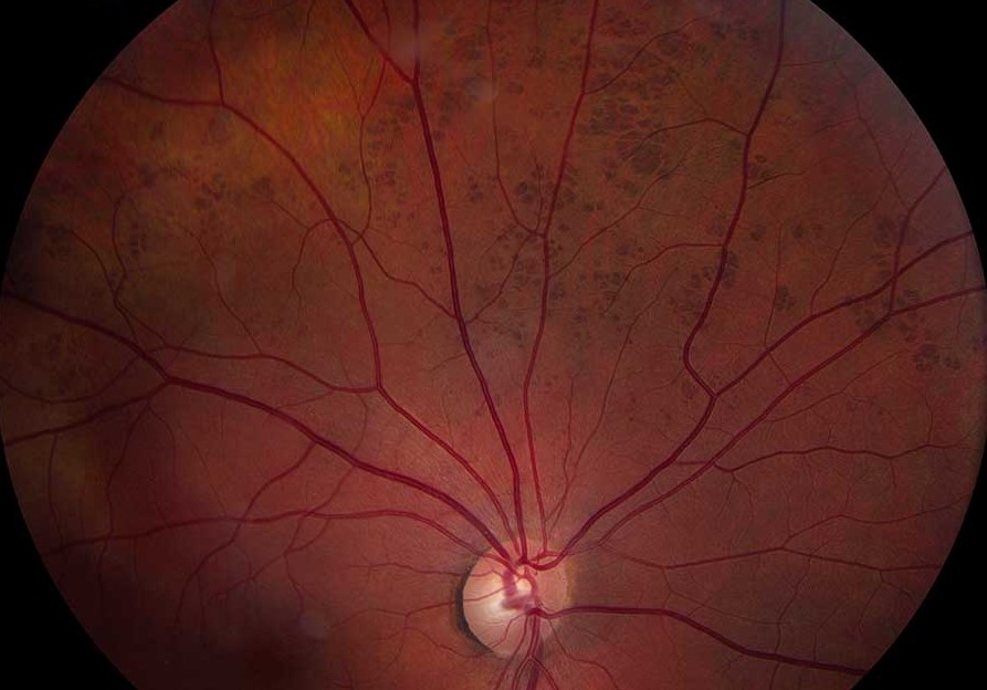
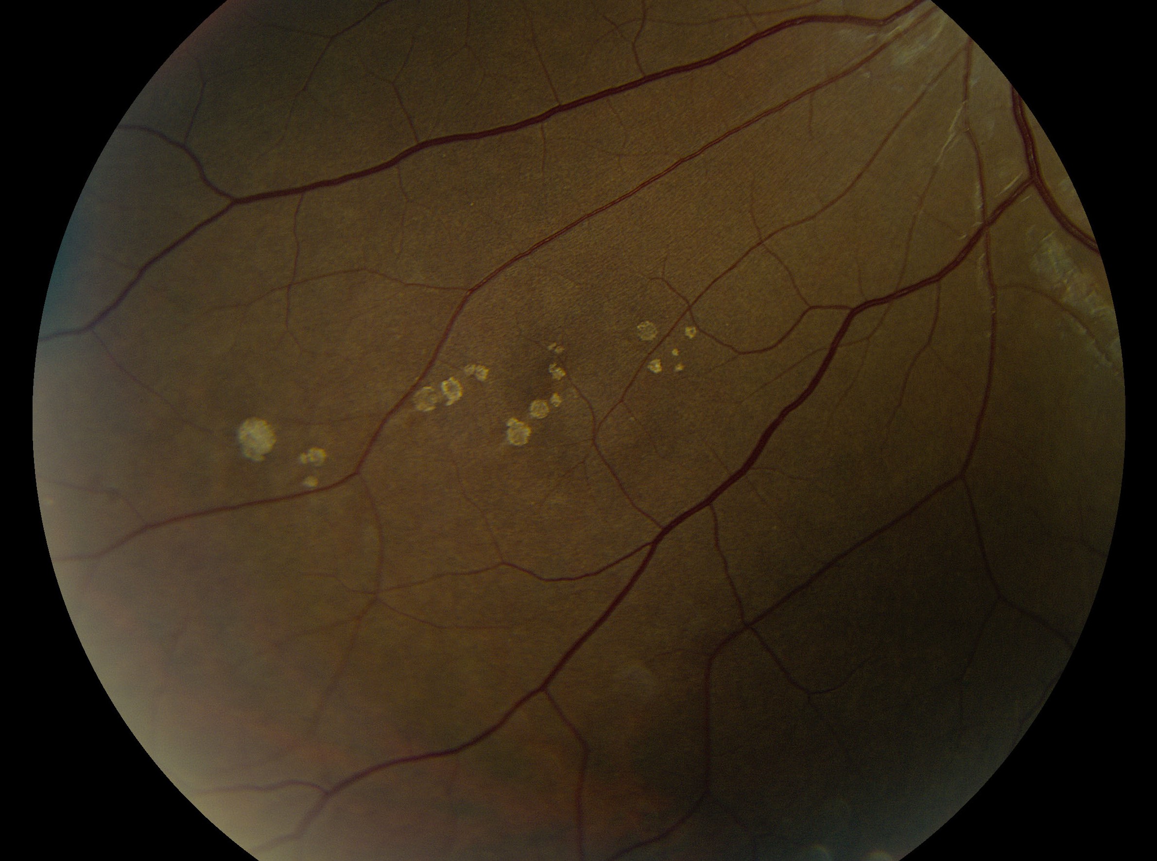
Bear tracks are clusters of flat, dark fundus spots caused by congenital grouped pigmentation of the retinal pigment epithelium (RPE).
Clinicians also call this pattern grouped CHRPE.
Most cases are incidental and benign.
However, a safe diagnosis requires the typical pattern and exclusion of its mimics.
What Bear Tracks Look Like
The spots resemble an animal’s paw prints, which explains the name.
Each lesion looks flat, sharply outlined and gray-brown to black.
Spots usually gather in one fundus sector.
In addition, they often enlarge toward the periphery.
Why the Grouped Pattern Forms
Histology shows RPE cells packed with large, round melanin granules.
Because the abnormality sits in the RPE, the overlying retina keeps its normal contour.
Meyer and colleagues linked the pattern to pigmentary mosaicism, although the mechanism remains uncertain.
Fundus Explorer Pro
Photograph the retinal findings described here with the phone already in your pocket — 22 D optics and built-in illumination in one handheld unit.
From Choroida — the team behind this siteSymptoms in Bear Tracks
Most patients have no symptoms and normal central vision.
Therefore, the finding usually surfaces during a routine dilated exam.
New photopsia or reduced vision should prompt a search for another cause.
Examining the Fundus
First, use indirect ophthalmoscopy to view the full sector.
Next, use a slit lamp lens to assess elevation, margins and surface changes.
Finally, examine the fellow eye, because bilateral disease widens the differential.
Imaging Bear Tracks
Widefield color photography provides the best baseline record.
OCT confirms a flat RPE-level lesion without subretinal fluid or a choroidal mass.
Pigmented lesions also tend to appear dark on fundus autofluorescence, although reports remain limited.
Fluorescein angiography and ultrasound add little when the appearance is classic.
Bear Tracks Versus Solitary CHRPE
Solitary lesions usually form one larger, round patch, often with a pale halo and lacunae.
Grouped lesions, by contrast, form many small spots without these features.
Shields and colleagues documented slow enlargement in many solitary lesions (see solitary CHRPE).
As a result, follow-up needs may differ between variants.
Polar Bear Tracks
A pale variant shows the same grouped arrangement with little or no pigment.
Clinicians often call these lesions polar bear tracks or grouped albinotic spots.
Published data remain limited, but most descriptions suggest an incidental finding.
Bear Tracks and Polyposis
Familial adenomatous polyposis can produce pigmented fundus lesions.
However, those lesions tend to be multiple, bilateral and widely scattered.
They are often oval or irregular, sometimes with a depigmented tail.
Traboulsi and colleagues described their value as a marker in Gardner syndrome.
In contrast, Shields and colleagues found no association between typical CHRPE and polyposis.
Therefore, take a family history and refer when the pattern or history fits.
Excluding Nevus and Melanoma
A choroidal nevus lies beneath the RPE and often has feathery margins or drusen.
Melanoma typically shows thickness, orange pigment, subretinal fluid or documented growth.
Because grouped lesions are flat and multiple, they rarely resemble melanoma.
Dense black lesions at the disc suggest choroidal melanocytoma, which needs separate assessment.
Other Lesions That Mimic Bear Tracks
- Reactive RPE hyperplasia: irregular pigment after trauma or inflammation
- Healed chorioretinitis: atrophic center with a pigmented border
- Retinitis pigmentosa: bone spicule pigment, night blindness and field loss
Findings That Need Workup
- Any measurable elevation or a dome-shaped lesion
- Orange pigment or subretinal fluid
- Documented growth or a new nodule
- Visual symptoms that the lesion cannot explain
- Numerous bilateral lesions or a family history of colorectal cancer
Managing Bear Tracks
Typical lesions need no laser, injection or surgery.
Treatment cannot remove the pigment, and it would add avoidable risk.
Management therefore centers on accurate classification and baseline photographs.
No trial defines a surveillance interval, so many clinicians check the lesion at routine dilated exams.
Then, refer to ocular oncology if elevation, fluid or growth appears.
Counseling Patients With Bear Tracks
Explain that these spots are usually congenital pigment clusters that rarely affect vision.
Next, advise the patient to report new flashes, floaters or visual loss.
Share the photographs, because a visual record often eases anxiety.
Pitfalls to Avoid
- Do not label a single large lesion as grouped CHRPE
- Avoid diagnosing from a poor peripheral view
- Do not skip the fellow eye
- Never ignore subretinal fluid because nearby spots look typical
- Do not order a polyposis workup from the classic sectoral pattern alone


Document what you see
Two smartphone imaging tools built for everyday clinic use — one for the slit lamp, one for the fundus.
From Choroida — the team behind this siteReferences
- Regillo CD, Eagle RC Jr, Shields JA, Shields CL, Arbizo VV. Histopathologic findings in congenital grouped pigmentation of the retina. Ophthalmology. 1993;100:400-405.
- Meyer CH, Rodrigues EB, Mennel S, Schmidt JC, Kroll P. Grouped congenital hypertrophy of the retinal pigment epithelium follows developmental patterns of pigmentary mosaicism. Ophthalmology. 2005;112:841-847.
- Shields CL, Mashayekhi A, Ho T, Cater J, Shields JA. Solitary congenital hypertrophy of the retinal pigment epithelium: clinical features and frequency of enlargement in 330 patients. Ophthalmology. 2003;110:1968-1976.
- Traboulsi EI, Krush AJ, Gardner EJ, et al. Prevalence and importance of pigmented ocular fundus lesions in Gardner’s syndrome. N Engl J Med. 1987;316:661-667.
- Shields JA, Shields CL, Shah PG, Pastore DJ, Imperiale SM Jr. Lack of association among typical congenital hypertrophy of the retinal pigment epithelium, adenomatous polyposis, and Gardner syndrome. Ophthalmology. 1992;99:1709-1713.

Writes and reviews the clinical articles on Educate. Every post is checked against peer-reviewed sources before it goes live.