
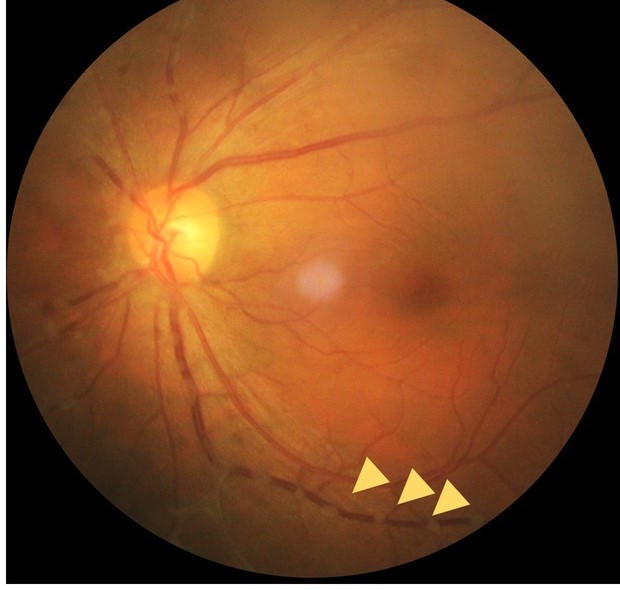
Chikungunya ocular manifestations are inflammatory eye complications of an alphavirus infection transmitted by Aedes mosquitoes.
The acute illness is characterized by fever and severe joint pain, and the eye disease usually appears in the second to third week or later.
Most ocular problems are self-limiting, but some cases involve the retina or optic nerve and need treatment.
What Is Chikungunya?
Chikungunya virus has caused outbreaks across Africa, Asia, the Indian Ocean, and the Americas.
The name refers to the stooped posture of patients with painful joints.
The illness starts with abrupt fever, polyarthralgia, rash, and headache, and the joint pain may last for months.
Ocular involvement is reported in a minority of patients, and it can occur even when the systemic illness was mild.
Chikungunya Ocular Manifestations
- Anterior uveitis: the commonest eye finding, granulomatous or nongranulomatous, with keratic precipitates and cells
- Episcleritis and conjunctivitis: early and often mild
- Optic neuritis and neuroretinitis: blurred vision, a relative afferent pupillary defect, and disc swelling (see optic neuritis)
- Retinitis and retinal vasculitis: with white patches and hemorrhages
- Macular edema and serous detachment: in a few patients
- Dry eye and keratitis: described
Ocular symptoms include redness, pain, blurred vision, floaters, and photophobia.
Most patients with anterior uveitis recover within weeks.
All-fit Slit-Lamp Adapter
Record and share exactly what you see at the slit lamp. One adapter fits any slit lamp or surgical microscope — and any smartphone.
From Choroida — the team behind this siteDifferential Diagnosis
- Dengue, Zika, and West Nile virus infections: other arboviruses that can cause retinal and optic nerve disease (see Zika virus retinopathy and West Nile retinopathy)
- Behçet disease, sarcoidosis, and HLA-B27 uveitis: noninfectious causes
- Infectious causes of uveitis: syphilis and tuberculosis
Epidemiology and exposure history help to separate these, since different viruses co-circulate in some regions.
Diagnosis
Diagnosis of the viral infection relies on RT-PCR in the first week, and IgM serology afterward.
Eye findings are diagnosed by examination, with OCT and, if needed, fluorescein angiography.
Intraocular fluid PCR is rarely required, but it may be considered in an atypical case.
Treatment
There is no specific antiviral treatment.
Topical corticosteroids and cycloplegics treat anterior uveitis, and the dose is tapered according to the response.
Systemic corticosteroids are used in optic neuritis, severe retinitis, or posterior uveitis, and the course is guided by the severity.
Monitor the pressure, the lens, and the macula during treatment, and review the patient until the inflammation has been quiet for some time.
Prevention is by controlling mosquitoes and by personal protection, including repellents and long clothing.
Patients with a history of chikungunya who develop eye symptoms should be examined promptly, since the timing may be late and the connection missed.
Timing and Course
Eye findings often arise in the subacute phase, between one and three weeks after the fever, although some cases are described months later.
Patients with persistent joint disease may have recurrent episodes of uveitis, and the symptoms can be attributed to the arthritis unless the eye is examined.
The inflammation of the anterior segment is often unilateral at the start and may become bilateral.
Retinitis and Neuroretinitis
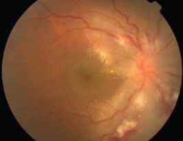
Retinitis appears as one or more areas of white retinal opacification with overlying vitreous cells, usually in the posterior pole.
Neuroretinitis is characterized by disc swelling and a macular star of exudates, and it may follow a mild course.
Optical coherence tomography and fluorescein angiography help to document the inflammation and to monitor the response (see fluorescein angiography patterns).
Visual field tests, such as the perimetry of the central field, show scotomas that match the lesions.
Management of Optic Neuritis
Optic neuritis from chikungunya is typically treated like other postinfectious optic neuritis, with intravenous methylprednisolone followed by an oral taper in severe cases.
Many patients recover vision, though some retain a residual defect in the visual field or color vision.
Check the visual acuity, color vision, and the visual field at follow-up, and repeat the OCT of the nerve fiber layer.
Counselling and Prevention
Patients in outbreak regions should know that eye symptoms can appear after the fever has resolved.
Advise them to seek an eye examination for pain, redness, blur, or floaters, and not to wait.
Public health measures to control the mosquito vector, including removal of standing water and the use of repellents, reduce the risk for communities.
The vaccine against chikungunya, where available, may offer protection for travelers and residents of outbreak areas.
Follow-Up
Examine the patient at intervals of one to two weeks while the inflammation is active, and then monthly until quiet.
A few patients have recurrences, particularly when joint symptoms flare, and they should be told to return.
Measure the pressure at each visit when steroids are used, and plan cataract assessment if a lens opacity develops.
Document the visual field and color vision in optic neuritis, because they are the best markers of recovery.


Document what you see
Two smartphone imaging tools built for everyday clinic use — one for the slit lamp, one for the fundus.
From Choroida — the team behind this siteReferences
- Mahendradas P, Ranganna SK, Shetty R, et al. Ocular manifestations associated with chikungunya. Ophthalmology. 2008;115:287-291.
- Mahendradas P, Avadhani K, Shetty R. Chikungunya and the eye: a review. J Ophthalmic Inflamm Infect. 2013;3:35.
- Salmon JF. Kanski’s Clinical Ophthalmology: A Systematic Approach. 9th ed. Elsevier; 2020.
- American Academy of Ophthalmology. Basic and Clinical Science Course, Section 9: Uveitis and Ocular Inflammation. AAO; current edition.

Writes and reviews the clinical articles on Educate. Every post is checked against peer-reviewed sources before it goes live.


