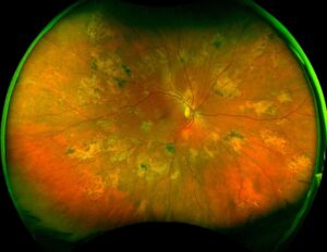
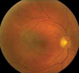
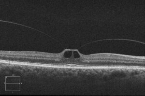
Quinine retinal toxicity produces sudden, severe, bilateral vision loss after a large dose of the drug.
It usually follows a deliberate overdose, but toxic levels can also occur with therapeutic misuse.
Because the early signs are dramatic, the diagnosis rests on the history.
Clinical Course of Quinine Retinal Toxicity
Patients first report profound loss of vision, and some are completely blind.
Also, the pupils are dilated and unreactive, and the fundus may look nearly normal early on.
Within days, central vision often recovers, but the visual field remains constricted.
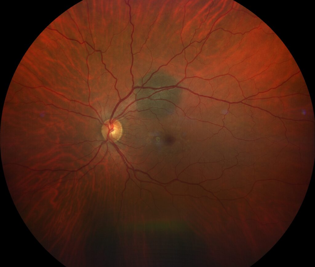
Late signs include narrowed retinal arterioles and optic disc pallor.
How Quinine Retinal Toxicity Develops
Brinton and colleagues proposed that vasoconstriction drives the injury.
Bacon and colleagues found evidence of a direct toxic effect on retinal cells.
As a result, the electroretinogram shows a reduced b-wave early on, and the amplitude may remain low.
Doses above about four grams are considered dangerous.
Fundus Explorer Pro
Photograph the retinal findings described here with the phone already in your pocket — 22 D optics and built-in illumination in one handheld unit.
From Choroida — the team behind this siteSystemic Features With Quinine Retinal Toxicity
- Tinnitus and hearing loss
- Nausea, vomiting, and headache
- Cardiac conduction abnormalities and hypotension
Therefore, the patient needs urgent toxicology and cardiac care.
Dyson and colleagues reported deaths from overdose, so the eye problem is secondary to the emergency.
Management of Quinine Retinal Toxicity
First, involve a poison control center and stabilize the patient.
Then document the field and the electroretinogram.
No treatment has proven benefit for the eye, and stellate ganglion block and hemodilution are not supported by evidence.
Finally, counsel patients that field loss is often permanent.
Differential Diagnosis of Sudden Bilateral Vision Loss
Quinine is one of several toxic causes of sudden visual loss in both eyes.
Consider methanol poisoning, which also causes dilated pupils and disc swelling.
Also think of bilateral optic neuritis, cortical blindness, and bilateral arterial occlusion.
In these conditions, the history and the pupil response guide the diagnosis.
Take a medication list for every patient, and ask about any intentional ingestion.
Prevention of Quinine Retinal Toxicity
Regulators advise against quinine for nocturnal leg cramps because the risks outweigh the benefit.
Therefore, review the medication list of patients with unexplained visual symptoms.
Also store the drug safely in homes where depression or self-harm is a concern.
The small amounts in tonic water are not a concern for the retina.
Visual Field and Electroretinogram in Quinine Retinal Toxicity
Fields show marked constriction, and a small central island often remains.
The electroretinogram shows a reduced b-wave, and in severe cases the a-wave declines too.
Bacon and colleagues found that the amplitudes may recover only partly.
Therefore, repeat fields at three and six months to document the final state.
Quinine Overdose Care and the Eye
Boland and colleagues described the systemic complications of poisoning.
Early gastric lavage and activated charcoal reduce absorption when given soon after ingestion.
Also, the eye team should stay involved, because vision often improves over days.
Document the pupil response daily, since return of reactivity is a good sign.
Finally, refer patients with suicidal intent for psychiatric assessment.
Pathophysiology in More Detail
Quinine affects the retinal ganglion cells and the photoreceptors, and it may also reduce blood flow.
Early reports described marked arteriolar narrowing, which suggested a vascular cause.
However, later studies showed that vessels may look normal early, so a direct toxic effect on the retina is likely.
Therefore, the injury probably has more than one mechanism.
Practical Points for the Clinician
Take a careful history of any ingested drugs, and ask relatives when the patient cannot answer.
Also check the electrocardiogram, since quinine prolongs the QT interval.
Then examine the pupils, the fundus, and the field, and repeat them over the first week.
Because many patients recover partly, give a cautious but hopeful message.
Long-Term Outlook
Central acuity often returns to normal or near normal.
However, the field remains constricted, and night vision is poor in many patients.
Therefore, refer for low vision rehabilitation when the field loss limits daily life.
Key Points for the Clinician
Sudden bilateral blindness after a drug ingestion should prompt a question about quinine.
Also, the pupils are dilated and fixed early, but vision may return.
Because the systemic risks are serious, work with toxicology and cardiology.
Then follow the field and the electroretinogram to document the outcome.
Finally, counsel patients and families about prevention of repeat exposure.
Counselling the Family
Explain that vision may improve, but that the field may stay small.
Also describe the signs of recovery, such as pupil reactivity.
Because the event often follows distress, offer support and referral.
Finally, share a plan for follow-up visits.
For the latest studies, see the PubMed results for quinine retinal toxicity.


Document what you see
Two smartphone imaging tools built for everyday clinic use — one for the slit lamp, one for the fundus.
From Choroida — the team behind this siteReferences
- Brinton GS, Norton EWD, Zahn JR, Levy IS. Ocular quinine toxicity. Am J Ophthalmol. 1980;90:403-410.
- Bacon P, Spalton DJ, Smith SE. Blindness from quinine toxicity. Br J Ophthalmol. 1988;72:219-224.
- Dyson EH, Proudfoot AT, Prescott LF, Heyworth R. Death and blindness due to overdose of quinine. Br Med J. 1985;291:31-33.
- Boland ME, Roper SM, Henry JA. Complications of quinine poisoning. Lancet. 1985;1:384-385.

Writes and reviews the clinical articles on Educate. Every post is checked against peer-reviewed sources before it goes live.