Intrachoroidal cavitation is a fluid-filled space inside the choroid, usually at the inferior edge of a highly myopic optic disc.
Early reports called it peripapillary detachment in pathologic myopia, but later OCT work showed that the space lies within the choroid itself. Therefore, the newer name describes its true location.
Who Develops Intrachoroidal Cavitation
The lesion appears mainly in eyes with high axial myopia. Most affected eyes also show a tilted disc and a wide myopic conus, and many have a posterior staphyloma or advanced myopic maculopathy. Population studies have also detected it in older adults with long eyes.
The Yellow-Orange Crescent
On fundus examination, look just beyond the inferior border of the conus, where a well-demarcated yellow-orange zone sits next to the disc. Its color is warmer than the white sclera inside the crescent (see myopic crescent). Because the change is subtle, it is easy to miss, although wide-field color photos often show it more clearly.
Fundus Explorer Pro
Photograph the retinal findings described here with the phone already in your pocket — 22 D optics and built-in illumination in one handheld unit.
From Choroida — the team behind this siteWhy the Sign Appears
The overlying retina and RPE stay near their normal level, while the sclera beneath bows backward and a space opens within the choroid. Hyporeflective fluid then fills the widened gap, but the exact mechanism is still debated.
OCT Signature of Intrachoroidal Cavitation
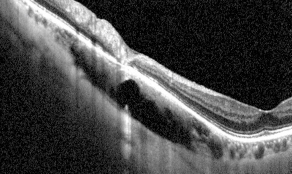
Enhanced depth imaging or swept-source OCT gives the best view, and the scan shows a wedge-shaped hyporeflective space inside the choroid. Above it, the RPE line runs level with the adjacent retina, while below it the scleral border dips posteriorly.
Communication With the Vitreous
Spaide and colleagues described tissue defects at the border of the conus in many eyes, and through these gaps the cavity may connect with the vitreous. Full-thickness retinal defects can also overlie the cavitation edge, and these openings may explain how fluid enters the choroid.
Vessel Kinking Over Intrachoroidal Cavitation
Retinal vessels crossing the lesion edge may bend sharply, and this step marks the transition from conus to cavitation. Therefore, a vessel kink at the inferior disc margin should prompt an OCT scan.
Visual Field Loss in Intrachoroidal Cavitation
Many patients show field defects that correspond to the lesion, and because the cavity sits inferiorly, the defect usually appears superiorly. These scotomas can look arcuate or paracentral, so they easily suggest glaucoma (see visual field trend analysis). Proposed causes include nerve fiber stretching and retinal defects at the cavity edge.
Intrachoroidal Cavitation or Glaucoma?
High myopia raises glaucoma risk, so both conditions may coexist. However, a defect that maps exactly onto the cavity points toward the lesion, although you should still check pressure, gonioscopy and rim configuration as usual. Normative RNFL databases perform poorly in tilted myopic discs, so interpret color-coded maps with caution.
Telling It From a Staphyloma
In a staphyloma, the retina and RPE follow the sclera backward, whereas in intrachoroidal cavitation the RPE stays in place while the sclera recedes. Therefore, an OCT gap between RPE and sclera separates the two (see peripapillary staphyloma). Clinically, a staphyloma shows a visible excavation with vessels dipping into it.
Other Mimics
- Optic disc pit: a gray oval depression within the disc, often with macular fluid
- Myopic conus: bare white sclera without a choroidal space
- Optic disc coloboma: an inferior excavation from incomplete fissure closure
- Choroidal mass: altered internal reflectivity, with no clear cavity
Natural History
Most lesions stay stable over years of follow-up, but some may enlarge slowly as the eye elongates. Ohno-Matsui and colleagues also described similar cavitation under the macula in pathologic myopia, so follow-up should include macular OCT as well as the disc.
Management of Intrachoroidal Cavitation
No treatment closes the cavity or restores the lost field, so management focuses on correct recognition and avoiding unnecessary glaucoma therapy. However, treat coexisting glaucoma when pressure or documented progression supports it, although pressure lowering will not reverse the cavitation itself.
Follow-Up Plan
- First, record a baseline color photo, OCT and visual field
- Then repeat fields to confirm stability
- Also check pressure at each visit
- Finally, scan the macula for schisis or new fluid
Counseling in Intrachoroidal Cavitation
Explain that the finding reflects the structure of a long myopic eye. Many patients never notice the field defect, and regular checks look for change and for glaucoma, while a stable lesion needs no procedure.
Pitfalls to Avoid
- Do not label a stable superior defect as glaucoma before scanning for cavitation
- Avoid reading RNFL maps in tilted discs at face value
- Never mistake the yellow-orange zone for a choroidal mass
- Remember macular OCT at the same visit


Document what you see
Two smartphone imaging tools built for everyday clinic use — one for the slit lamp, one for the fundus.
From Choroida — the team behind this siteReferences
- Freund KB, Ciardella AP, Yannuzzi LA, et al. Peripapillary detachment in pathologic myopia. Arch Ophthalmol. 2003;121:197-204.
- Toranzo J, Cohen SY, Erginay A, Gaudric A. Peripapillary intrachoroidal cavitation in myopia. Am J Ophthalmol. 2005;140:731-732.
- Shimada N, Ohno-Matsui K, Yoshida T, et al. Characteristics of peripapillary detachment in pathologic myopia. Arch Ophthalmol. 2006;124:46-52.
- Spaide RF, Akiba M, Ohno-Matsui K. Evaluation of peripapillary intrachoroidal cavitation with swept source and enhanced depth imaging optical coherence tomography. Retina. 2012;32:1037-1044.
- Ohno-Matsui K, Akiba M, Moriyama M, et al. Intrachoroidal cavitation in macular area of eyes with pathologic myopia. Am J Ophthalmol. 2012;154:382-393.
Test yourself
A few questions straight from this article.
-
Where does peripapillary intrachoroidal cavitation usually sit relative to the optic disc?
It is a fluid-filled space inside the choroid, usually at the inferior edge of a highly myopic optic disc, often with a tilted disc and wide conus. -
Which fundus sign suggests peripapillary intrachoroidal cavitation in a highly myopic eye?
A well-demarcated yellow-orange zone sits just beyond the inferior conus, warmer in colour than the white sclera inside the crescent; it is subtle and easily missed. -
What does OCT show in peripapillary intrachoroidal cavitation?
The RPE line runs level with the adjacent retina while the scleral border beneath dips posteriorly, leaving a wedge-shaped hyporeflective choroidal space. -
Which OCT techniques give the best view of peripapillary intrachoroidal cavitation?
Enhanced depth imaging or swept-source OCT gives the best view of the choroidal space and the posteriorly bowed sclera. -
What did Spaide and colleagues describe at the conus border in intrachoroidal cavitation?
Tissue defects at the conus border may connect the cavity with the vitreous, and these openings may explain how fluid enters the choroid. -
In a highly myopic eye, a retinal vessel kinking sharply at the inferior disc margin should prompt what?
A vessel bending sharply at the lesion edge marks the step from conus to cavitation, so it should prompt an OCT scan. -
Where does the visual field defect from peripapillary intrachoroidal cavitation usually appear?
Because the cavity sits inferiorly, the defect usually appears superiorly and may look arcuate or paracentral, easily suggesting glaucoma. -
On OCT, what separates peripapillary intrachoroidal cavitation from a peripapillary staphyloma?
In a staphyloma the retina and RPE follow the sclera backward, whereas in cavitation the RPE stays in place, leaving an RPE-sclera gap. -
What is the main management goal in peripapillary intrachoroidal cavitation?
No treatment closes the cavity or restores the field; coexisting glaucoma is treated when pressure or documented progression supports it. -
Why should follow-up of peripapillary intrachoroidal cavitation include macular OCT?
Ohno-Matsui and colleagues described similar cavitation under the macula in pathologic myopia, and the macula should also be checked for schisis or new fluid.

Writes and reviews the clinical articles on Educate. Every post is checked against peer-reviewed sources before it goes live.