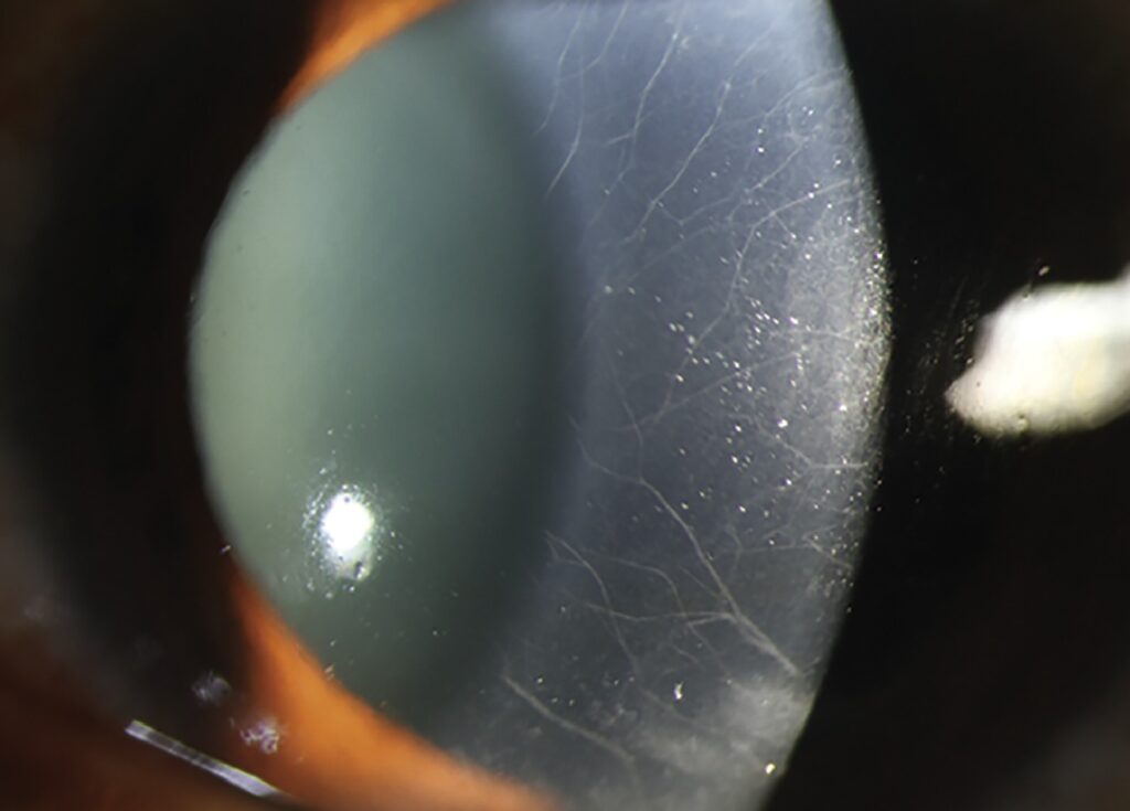
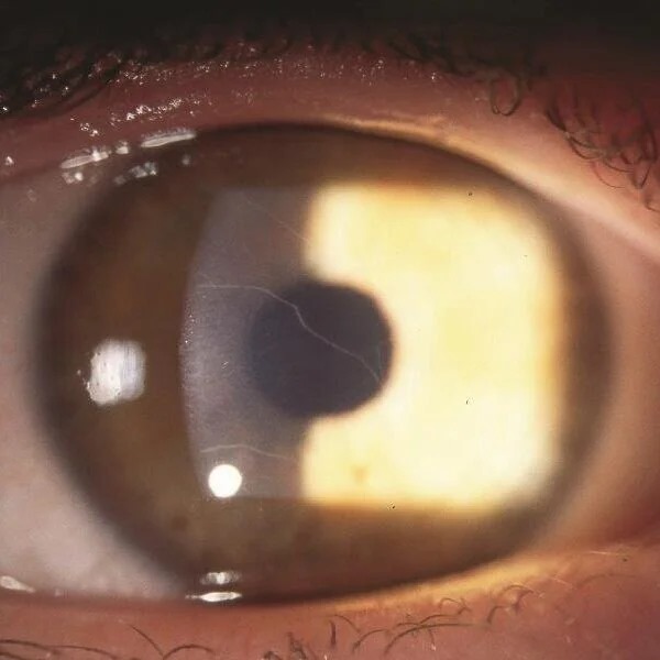
Prominent corneal nerves are stromal nerve fibers that look thicker, whiter or more numerous than normal at the slit lamp.
Many healthy eyes show some nerves, mostly in the peripheral and mid stroma. However, nerves that cross the central cornea or look beaded deserve a structured workup. Most causes are local, while one is a warning sign of thyroid cancer.
What Normal Corneal Nerves Look Like
Corneal nerves enter the stroma at the limbus as branches of the long ciliary nerves, and they run radially toward the center and branch as they become more superficial. In adults, the peripheral trunks often appear as fine white lines. As a result, a few visible nerves near the limbus are usually normal.
How to Examine Prominent Corneal Nerves
Start with direct focal illumination and a narrow slit at moderate magnification, then use retroillumination from the iris, which shows nerves as fine refractile threads. Sclerotic scatter also helps because it outlines the whole network at once, and for technique, review slit lamp illumination methods.
All-fit Slit-Lamp Adapter
Record and share exactly what you see at the slit lamp. One adapter fits any slit lamp or surgical microscope — and any smartphone.
From Choroida — the team behind this siteSigns That Make Nerves Abnormal
Abnormal nerves look thick, beaded, or extend well into the central cornea, so also note whether infiltrates surround them. Finally, compare both eyes, because systemic causes usually produce bilateral, symmetric changes.
MEN 2B and Prominent Corneal Nerves
Multiple endocrine neoplasia type 2B is the most important cause to recognize, and it results from activating mutations in the RET proto-oncogene. The nerves are markedly thickened, bilateral and often cross the pupillary zone, yet patients usually keep normal vision and report few eye symptoms. Therefore, the eye examiner may be the first to suspect the syndrome.
Systemic Clues in MEN 2B
Look beyond the cornea when a young patient shows these nerves.
- Mucosal neuromas on the lips, tongue or eyelid margins
- Thickened, slightly everted eyelids and full lips
- Marfanoid body habitus
- Bowel symptoms from intestinal ganglioneuromatosis
- Personal or family history of medullary thyroid carcinoma or pheochromocytoma
Urgent Referral in MEN 2B
Medullary thyroid carcinoma in MEN 2B tends to appear early and behave aggressively. Guidelines therefore advise genetic testing and very early prophylactic thyroidectomy in carriers. Refer to endocrinology promptly and offer testing to first-degree relatives, and exclude pheochromocytoma before any planned surgery.
Keratoconus and Prominent Corneal Nerves
In keratoconus, nerves often look more visible within the thinned stroma. Supporting signs include Vogt striae, a Fleischer ring and a scissoring reflex, and corneal topography then confirms the diagnosis. Also ask about eye rubbing, which may drive progression (see eye rubbing and keratoconus).
Fuchs Dystrophy
In Fuchs endothelial dystrophy, nerves may stand out against guttae and stromal edema. Specular reflection shows the guttae, while pachymetry tracks the edema. Because the nerves are a secondary finding, management targets the endothelium (see Fuchs endothelial corneal dystrophy).
Acanthamoeba Keratitis
Radial keratoneuritis is an early and highly suggestive sign of Acanthamoeba infection, and infiltrates line the nerves in a radial pattern. Pain is often severe and out of proportion to the findings, so ask every contact lens wearer about water exposure and lens hygiene. Arrange confocal microscopy or culture promptly, because early treatment improves outcomes.
Leprosy and Prominent Corneal Nerves
Hansen disease classically produces thickened, beaded corneal nerves, and Mycobacterium leprae has a tropism for peripheral nerves, including those of the cornea. Corneal sensation often falls, and lagophthalmos adds exposure risk. Look also for madarosis, punctate keratitis and iris pearls.
Other Causes of Prominent Corneal Nerves
Several less common conditions complete the differential.
- Neurofibromatosis type 1
- Ichthyosis
- Refsum disease
- Congenital glaucoma
- Posterior polymorphous corneal dystrophy
- Failed corneal graft or previous trauma
- Idiopathic finding or normal variant
Confocal Microscopy in Prominent Corneal Nerves
In vivo confocal microscopy shows nerve diameter, tortuosity and density at cellular resolution, and it can also reveal Acanthamoeba cysts in suspected keratitis. However, interpretation needs experience, and normal values differ between devices (for details, see corneal nerve imaging).
A Practical Workup
First, decide whether the nerves are truly abnormal or simply well seen, and next test corneal sensation and ask about contact lenses and pain. Then look for shape change, guttae and infiltrates, and finally, if both corneas show thick nerves without a local cause, examine the lips, tongue and neck.
Counseling Patients with Prominent Corneal Nerves
The nerves themselves rarely cause symptoms, but explain that the finding is a clue, so the cause decides treatment. When MEN 2B is possible, speak plainly about blood tests and genetic counseling, while for a normal variant, reassure the patient and record the finding.
Follow-Up of Prominent Corneal Nerves
Photograph the cornea at baseline so later changes are easy to compare. In infectious causes, review often until the infiltrates settle, and meanwhile repeat topography in keratoconus to detect progression.
Pitfalls to Avoid
- Do not label normal peripheral nerves as pathologic
- Never miss mucosal neuromas in a child with bilateral thick nerves
- Avoid treating radial keratoneuritis as herpes keratitis without testing
- Always examine the fellow eye


Document what you see
Two smartphone imaging tools built for everyday clinic use — one for the slit lamp, one for the fundus.
From Choroida — the team behind this siteReferences
- Bowling B. Kanski’s Clinical Ophthalmology: A Systematic Approach. 8th ed. Elsevier; 2016.
- American Academy of Ophthalmology. Basic and Clinical Science Course, Section 8: External Disease and Cornea. San Francisco: AAO; 2023.
- Wells SA Jr, Asa SL, Dralle H, et al. Revised American Thyroid Association guidelines for the management of medullary thyroid carcinoma. Thyroid. 2015;25:567-610.
- Dart JK, Saw VP, Kilvington S. Acanthamoeba keratitis: diagnosis and treatment update 2009. Am J Ophthalmol. 2009;148:487-499.
- Cruzat A, Qazi Y, Hamrah P. In vivo confocal microscopy of corneal nerves in health and disease. Ocul Surf. 2017;15:15-47.
Test yourself
A few questions straight from this article.
-
Which gene's activating mutations cause multiple endocrine neoplasia type 2B, the key systemic cause of prominent corneal nerves?
The article states that MEN 2B results from activating mutations in the RET proto-oncogene. It is the most important cause of prominent corneal nerves to recognize. -
What corneal nerve pattern is typical of MEN 2B in a patient who usually has normal vision?
In MEN 2B the nerves are markedly thickened, bilateral and often cross the pupillary zone. Patients usually keep normal vision, so the eye examiner may be the first to suspect the syndrome. -
Which systemic finding should prompt suspicion of MEN 2B in a young patient with prominent corneal nerves?
The article lists mucosal neuromas of the lips, tongue or eyelid margins among the systemic clues for MEN 2B, along with thickened everted eyelids and marfanoid habitus. -
What is the recommended management for carriers of MEN 2B, given the behavior of its medullary thyroid carcinoma?
Medullary thyroid carcinoma in MEN 2B appears early and behaves aggressively. Guidelines therefore advise genetic testing and very early prophylactic thyroidectomy in carriers. -
What should be excluded before any planned surgery in a patient with MEN 2B?
The article advises referring promptly to endocrinology, offering testing to first-degree relatives, and excluding pheochromocytoma before any planned surgery. -
Which signs support keratoconus when corneal nerves look more visible within a thinned stroma?
In keratoconus, supporting signs include Vogt striae, a Fleischer ring and a scissoring reflex. Corneal topography then confirms the diagnosis. -
Which early, highly suggestive sign of Acanthamoeba keratitis involves the corneal nerves?
Radial keratoneuritis is an early and highly suggestive sign of Acanthamoeba infection, with infiltrates lining the nerves in a radial pattern. -
What should be asked of every contact lens wearer with suspected Acanthamoeba keratitis?
Pain is often severe and out of proportion to the findings, so the article advises asking every contact lens wearer about water exposure and lens hygiene. -
Which corneal nerve findings classically occur in leprosy (Hansen disease) due to Mycobacterium leprae?
Hansen disease classically produces thickened, beaded corneal nerves because M. leprae has a tropism for peripheral nerves. Corneal sensation often falls, and lagophthalmos adds exposure risk. -
In Fuchs endothelial dystrophy with visible corneal nerves, what is the management focus?
In Fuchs dystrophy the nerves may stand out against guttae and edema, but they are secondary, so management targets the endothelium.

Writes and reviews the clinical articles on Educate. Every post is checked against peer-reviewed sources before it goes live.