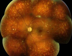
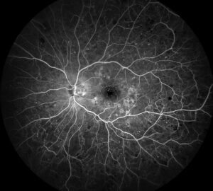
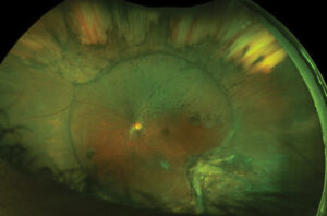
Deferoxamine retinopathy is a toxic retinal disorder that can occur in patients receiving iron chelation with deferoxamine, also called desferrioxamine.
The patients are usually those with thalassemia or other transfusion-dependent anemias.
The retinal damage can reduce vision if it goes unrecognized.
Regular monitoring and attention to symptoms allow early recognition and often recovery.
Why Deferoxamine Retinopathy Occurs
Patients who receive regular transfusions accumulate iron, and chelators bind the metal and promote its excretion.
Also, clinicians give deferoxamine by slow subcutaneous or intravenous infusion.
Its side effects include effects on hearing, vision, and growth.
The retinal pigment epithelium is also rich in metabolically active cells and iron-dependent enzymes.
It appears to be sensitive to the drug.
Toxicity is more likely with high doses, with high dose relative to the iron burden, and in patients with low iron stores, in whom the drug may also bind essential metals.
Clinical Features of Deferoxamine Retinopathy
- Visual symptoms: reduced night vision, loss of color vision, reduced acuity, and visual field loss, sometimes acute after a high dose or a pulse of treatment
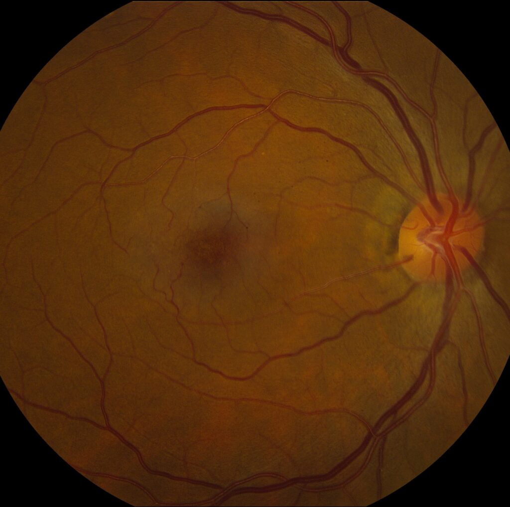
- Fundus findings: often subtle in the early stage, with later pigment stippling, a macular pattern resembling a pattern dystrophy, and RPE atrophy
- Electrophysiology: abnormal electro-oculogram and ERG with reduced rod and cone function, in many cases
- Visual fields: central or paracentral scotomas, and constriction
- Associated problems: hearing loss and, rarely, optic neuropathy
The first reports described acute visual loss after high-dose intravenous therapy, and later reports described a spectrum of chronic changes (see the references).
Fundus Explorer Pro
Photograph the retinal findings described here with the phone already in your pocket — 22 D optics and built-in illumination in one handheld unit.
From Choroida — the team behind this siteRisk Factors of Deferoxamine Retinopathy
- High daily dose and rapid infusion
- Low serum ferritin or a high ratio of dose to iron load
- Renal impairment, which impairs clearance of the drug
- Young age and long duration of treatment
- Concomitant diseases, such as diabetes
Modern dosing regimens aim to avoid excessive doses, and the risk is lower than in the early years.
Differential Diagnosis of Deferoxamine Retinopathy
- Thalassemia retinopathy itself, with pigment changes and angioid-like streaks (see thalassemia retinopathy)
- Other drug toxicity, such as hydroxychloroquine and thioridazine (see thioridazine retinopathy)
- Inherited pattern dystrophies and retinitis pigmentosa (see pattern dystrophies)
- Vitamin A deficiency and other metabolic disorders (see Bitot spots and vitamin A deficiency)
- Optic neuropathy and other visual pathway problems, in patients with visual field loss
A review of the medications, and the timing of symptoms in relation to treatment, support the diagnosis.
Monitoring
- Baseline eye examination before treatment, with acuity, color vision, fundus examination, and visual fields
- Regular examinations during treatment, such as yearly, or more often in patients at risk
- Electrophysiology when the findings are uncertain, or when toxicity is suspected
- Audiometry, since hearing loss can occur at the same time
- Prompt assessment of any new symptoms, such as night blindness or color changes
Hematology societies usually write monitoring guidelines, and ophthalmologists should know the local protocol.
Management
The key step is to stop or reduce the drug, and to discuss with the hematologist whether to use a different chelator.
Vision may recover in the weeks to months after cessation, particularly in patients who stop early, though some have permanent damage.
Oral chelators, such as deferasirox and deferiprone, are alternatives.
They have their own toxicity profiles, with ocular effects reported rarely.
The patient needs to continue chelation in some form, so the choice has to be made with a balance of risks.
Counselling
Explain to patients and families that the drug is necessary to prevent the harm of iron overload, but that the eyes need to be checked, and that symptoms should be reported quickly.
Provide written information, and coordinate follow-up with the hematology clinic.
Case Pattern
A young woman with thalassemia major who receives deferoxamine infusions at a high dose complains of difficulty seeing in dim light and notes that colors look washed out.
Her acuity is 20/30, and the fundus shows subtle mottling at the macula.
The ERG shows reduced responses, and the hematologist, informed of the findings, reduces the dose and considers switching to an oral chelator.
Over the following months her night vision and acuity recover, and the ERG improves.
Monitoring Intervals in Practice
Many protocols recommend an annual eye examination for patients on regular deferoxamine, and more frequent visits, such as every three to six months, for those on high doses, with low ferritin, or with symptoms.
Electrophysiology is useful as a baseline and when symptoms occur.
Keep a file with the dates, the dose, and the results, and share it with the hematology clinic.
Other Chelators and the Eye
Deferiprone and deferasirox have been associated with rare ocular side effects, such as lens opacities and retinal changes, so patients on these drugs should also be monitored.
The specific monitoring recommendations differ between drugs, so consult the prescribing information.
For the latest studies, see the PubMed results for deferoxamine retinopathy.


Document what you see
Two smartphone imaging tools built for everyday clinic use — one for the slit lamp, one for the fundus.
From Choroida — the team behind this siteReferences
- Davies SC, Marcus RE, Hungerford JL, et al. Ocular toxicity of high-dose intravenous desferrioxamine. Lancet. 1983;2:181-184.
- Lakhanpal V, Schocket SS, Jiji R. Deferoxamine (Desferal)-induced toxic retinal pigmentary degeneration and presumed optic neuropathy. Ophthalmology. 1984;91:443-451.
- Olivieri NF, Buncic JR, Chew E, et al. Visual and auditory neurotoxicity in patients receiving subcutaneous deferoxamine infusions. N Engl J Med. 1986;314:869-873.
- American Academy of Ophthalmology. Basic and Clinical Science Course, Section 12: Retina and Vitreous. AAO; current edition.

Writes and reviews the clinical articles on Educate. Every post is checked against peer-reviewed sources before it goes live.