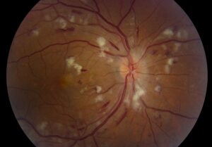
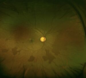
Peripheral retinal degenerations are age-related and developmental changes in the retina beyond the equator, and most are harmless.
A few raise the risk of retinal tears and detachment, and the clinical skill is also to tell them apart.
The same dilated examination that finds a dangerous lesion will also reveal many benign ones, and patients need reassurance as well as vigilance.
How to Approach Peripheral Retinal Degenerations
A careful examination with scleral depression is needed to see the periphery fully (see scleral depression).
In addition, the examiner looks for the type of lesion, the presence of breaks, vitreoretinal traction, and the status of the vitreous.
Risk is also judged by the lesion and by the patient: myopia, family history, history of detachment in the fellow eye, and previous eye surgery (see fellow eye retinal detachment risk).
Peripheral Retinal Degenerations: Lesions With Little or No Risk
- Paving stone (cobblestone) degeneration: discrete, round, yellow-white areas of chorioretinal atrophy, typically near the ora, which rarely cause breaks (see paving stone degeneration)
- Peripheral cystoid degeneration: microcysts near the ora serrata, found in nearly all adults, with a reticular pattern at times
- Pigmentary changes and perivascular pigment, often benign
- Ora serrata pearls and enclosed bays, variants of normal anatomy
- White without pressure and white with pressure: patches that appear white on indentation, mostly benign (see white without pressure)
- Retinal tufts of various types, which carry a small risk (see retinal tufts)
Patients with these lesions need no treatment, so routine care is sufficient.
Fundus Explorer Pro
Photograph the retinal findings described here with the phone already in your pocket — 22 D optics and built-in illumination in one handheld unit.
From Choroida — the team behind this siteLesions With Potential Risk
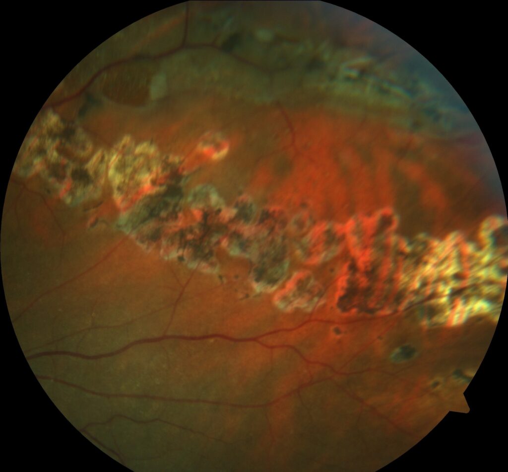
- Lattice degeneration: a thinning of the retina with overlying liquefied vitreous, with firm adhesion at the edges, which can also cause tears (see lattice degeneration)
- Snail track degeneration: a variant of lattice with glistening dots (see snail track degeneration)
- Degenerative retinoschisis: a splitting of the retinal layers, usually inferotemporally, which rarely progresses to detachment (see senile retinoschisis)
- Meridional folds and complexes with traction
- Zonular traction tufts and cystic tufts, which can also form tears
- Atrophic round holes, within lattice or alone (see operculated retinal hole)
Their risk, however, is a function of how they interact with the vitreous.
Peripheral Retinal Degenerations: What Raises the Risk
- Symptomatic posterior vitreous detachment, with new flashes and floaters
- Retinal tears with traction, especially horseshoe tears
- High myopia and a family history of detachment
- Previous detachment in the fellow eye
- Aphakia or pseudophakia, particularly after complicated surgery (see pseudophakic retinal detachment)
- Stickler syndrome and other connective tissue disorders (see Stickler syndrome)
- Trauma
Peripheral Retinal Degenerations: Management Principles
- Observation: for most asymptomatic lesions without breaks or traction, with education about symptoms
- Laser retinopexy or cryotherapy: for symptomatic tears, for high-risk breaks, and in selected patients with risk factors (see laser retinopexy and retinal cryotherapy)
- Prophylaxis of lattice without breaks: not routinely performed, since evidence is weak, but considered when the fellow eye has detached or in special risk groups
- Surgery: for retinal detachment, according to the type
A Cochrane review found that the evidence for prophylactic treatment of asymptomatic breaks and lattice is limited, so treatment decisions are individualized (see the references).
Patient Education
- First, explain that most peripheral changes are benign
- Then teach the warning symptoms: new flashes, a shower of floaters, a shadow, or a curtain in the vision
- Advise prompt examination if these occur, and routine reviews at an interval based on the risk
- Finally, document the findings with a drawing or a widefield image for comparison
Reassure patients that changes in the periphery are common and do not mean that the retina will detach.
Frequency in the Population
Peripheral degenerations are very common.
Also, lattice degeneration is found in about 6 to 10 percent of the population, and in a higher proportion of myopes and of patients with a detachment.
Also, peripheral cystoid degeneration is present in nearly all adults, and paving stone degeneration in about a quarter of adults over 20 in some series.
Because of this frequency, most lesions will never cause trouble, and detachments arise in a small proportion of those who have them.
Practical Documentation
Draw or photograph each clinically relevant lesion, with its location by clock hour and distance from the ora.
In addition, note any associated breaks, vitreous traction, and pigment, and the presence of a posterior vitreous detachment.
Use consistent colors on drawings, such as red for tears and holes, blue for detached retina, and brown or black for pigment, which aids communication.
Common Questions From Patients
- Will it detach? Most do not, and the risk can be monitored
- Should I avoid activities? There is no need to avoid normal activities, though contact sports carry the risk of trauma
- Can I have laser? It is considered in selected cases
- Do my children need examination? Offer it if there is a family history of detachment or a hereditary condition


Document what you see
Two smartphone imaging tools built for everyday clinic use — one for the slit lamp, one for the fundus.
From Choroida — the team behind this siteReferences
- Byer NE. Long-term natural history of lattice degeneration of the retina. Ophthalmology. 1989;96:1396-1402.
- Wilkinson CP. Interventions for asymptomatic retinal breaks and lattice degeneration for preventing retinal detachment. Cochrane Database Syst Rev. 2014;9:CD003170.
- Ryan SJ, Schachat AP, Wilkinson CP, et al, eds. Retina. 6th ed. Elsevier; 2018.
- American Academy of Ophthalmology. Basic and Clinical Science Course, Section 12: Retina and Vitreous. AAO; current edition.

Writes and reviews the clinical articles on Educate. Every post is checked against peer-reviewed sources before it goes live.