Intravitreal triamcinolone was the first widely used steroid injection for macular edema, and it remains a useful and inexpensive option.
It reduces edema quickly, but its effect wears off within a few months, and cataract and pressure rise are the price in many patients.
Understanding the trials helps to decide who should receive it.
What Is Intravitreal Triamcinolone?
Triamcinolone acetonide is a synthetic corticosteroid, and it is given as a crystalline suspension injected into the vitreous in a dose of about 1 to 4 milligrams.
Also, the crystals dissolve slowly, and the drug acts for about three months in many eyes.
It reduces inflammation, vascular permeability, and the production of VEGF, so it decreases leakage and swelling.
Clinicians prefer preservative-free preparations for intravitreal use, since the preservative in some products can cause toxicity.
Indications of Intravitreal Triamcinolone
- Diabetic macular edema, especially in pseudophakic eyes or when anti-VEGF drugs are not suitable (see diabetic macular edema)
- Macular edema after vein occlusion (see central retinal vein occlusion and branch retinal vein occlusion)
- Uveitic macular edema and inflammatory conditions (see uveitic macular edema)
- Pseudophakic cystoid macular edema that does not respond to drops (see Irvine-Gass syndrome)
- Visualization of the vitreous during vitrectomy, as a staining agent
- Neovascular glaucoma and other conditions, in selected cases
The use of triamcinolone for macular edema is off-label in many countries, with the exception of approved formulations.
Fundus Explorer Pro
Photograph the retinal findings described here with the phone already in your pocket — 22 D optics and built-in illumination in one handheld unit.
From Choroida — the team behind this siteWhat the Trials Showed
The Standard Care versus Corticosteroid for Retinal Vein Occlusion (SCORE) study compared 1 and 4 milligram doses with standard care.
In central vein occlusion, both doses improved vision more than observation, with similar benefit and a better safety profile for the 1 milligram dose (see the references).
In branch vein occlusion, triamcinolone was not better than grid laser in the study, so laser remained the standard at that time.
The DRCR network compared 1 and 4 milligram doses of triamcinolone with focal and grid laser for diabetic macular edema (see focal and grid laser).
Triamcinolone showed an early benefit in vision, but over two years laser was as good or better, and triamcinolone had more side effects, so laser became the preferred option at that time (see the references).
Since then, anti-VEGF drugs have replaced both as the main treatment.
Risks of Intravitreal Triamcinolone
- Raised intraocular pressure: in about a third or more of eyes, usually manageable with drops, but requiring laser or surgery in a few (see steroid-induced glaucoma)
- Cataract: common in phakic eyes, with progression over the first year or two
- Endophthalmitis: rare, as with any injection
- Sterile endophthalmitis, a noninfectious inflammation within days of injection, which may be mistaken for infection (see post-injection endophthalmitis)
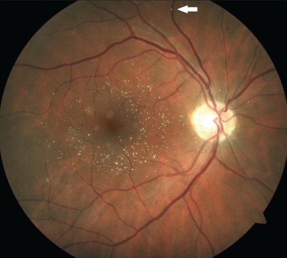
- Crystalline deposits and pseudohypopyon
- Vitreous hemorrhage, retinal detachment, and injection site complications
Patients should be monitored for pressure at one to two weeks and at intervals thereafter.
Practical Technique
- Use a preservative-free preparation, and a small gauge needle
- Place the injection as for other intravitreal drugs, with antisepsis, anesthesia, and a speculum (see intravitreal injection technique)
- Inject slowly and avoid reflux, since the crystals can be seen
- Check the pressure and the perfusion of the optic nerve after the injection
Many centers use triamcinolone in combination with other treatments, such as laser or anti-VEGF drugs.
Who Is a Good Candidate?
- Pseudophakic eyes, since cataract is not a concern
- Patients who cannot attend frequent visits for injections
- Eyes that respond poorly to anti-VEGF drugs
- Patients without a history of steroid response or glaucoma
Patients at high risk of raised pressure, or with advanced glaucoma, should be considered for other treatments.
Counselling
Explain that the injection can reduce swelling quickly, that the effect fades over months, and that cataract and raised pressure are likely.
In addition, tell the patient to attend for pressure checks, and to report pain, redness, or reduced vision.
Choosing Triamcinolone in Practice
For a pseudophakic patient with persistent diabetic edema after several anti-VEGF injections, a single steroid injection often gives a clear drop in thickness.
The result can guide the choice of a longer-acting implant.
For a phakic patient, discuss cataract surgery as a likely later step, and consider combining the plan with the timing of that operation.
Document the baseline pressure, repeat it after the injection, and record the response on OCT at four to six weeks, so that the decision to repeat is based on measured benefit.


Document what you see
Two smartphone imaging tools built for everyday clinic use — one for the slit lamp, one for the fundus.
From Choroida — the team behind this siteReferences
- SCORE Study Research Group. A randomized trial comparing the efficacy and safety of intravitreal triamcinolone with standard care to treat vision loss associated with macular edema secondary to central retinal vein occlusion: the Standard Care vs Corticosteroid for Retinal Vein Occlusion (SCORE) study report 5. Arch Ophthalmol. 2009;127:1101-1114.
- Diabetic Retinopathy Clinical Research Network. A randomized trial comparing intravitreal triamcinolone acetonide and focal/grid photocoagulation for diabetic macular edema. Ophthalmology. 2008;115:1447-1459.
- Ryan SJ, Schachat AP, Wilkinson CP, et al, eds. Retina. 6th ed. Elsevier; 2018.
- American Academy of Ophthalmology. Basic and Clinical Science Course, Section 12: Retina and Vitreous. AAO; current edition.

Writes and reviews the clinical articles on Educate. Every post is checked against peer-reviewed sources before it goes live.