
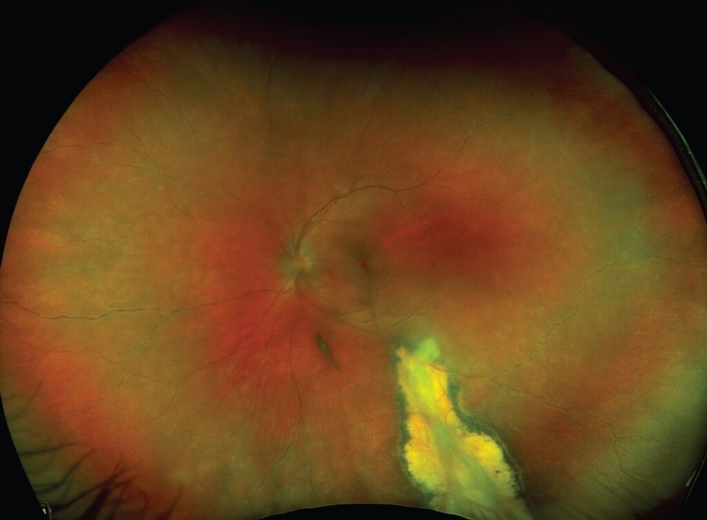
Ocular Lyme disease is the term for the eye findings of infection with Borrelia burgdorferi, the spirochete transmitted by Ixodes ticks.
The eye can be involved at any stage, from a mild conjunctivitis in the early phase to uveitis and optic neuropathy later on.
The ocular signs are not specific, so the diagnosis depends on exposure history, the pattern of findings, and serology.
What Is Lyme Disease, and What Is Ocular Lyme Disease?
Lyme disease occurs in the northern hemisphere in areas where the tick vector lives, and cases cluster in spring and summer.
The illness has three stages: early localized disease, with the erythema migrans rash, early disseminated disease, with facial palsy, meningitis, radiculitis, and carditis, and late disease, with arthritis and chronic neurological problems.
Many patients do not recall a tick bite, and the rash may be absent or missed.
The ophthalmologist often sees the patient because of a red eye or a visual disturbance that was not linked to the systemic infection.
Ocular Manifestations
- Follicular conjunctivitis: frequent in the early stage, mild and self-limited
- Episcleritis and keratitis: sometimes with stromal infiltrates that resemble interstitial keratitis (see interstitial keratitis)
- Uveitis: anterior, intermediate, and posterior forms, and sometimes a picture like pars planitis (see pars planitis)
- Retinal vasculitis and choroiditis
- Neuroretinitis and optic neuritis: with disc swelling and a macular star (see neuroretinitis and optic neuritis)
- Cranial neuropathies: facial and abducens palsies, and papilledema from raised intracranial pressure
In a patient with a facial palsy or abducens palsy in an endemic area, Lyme disease is an important cause, particularly in children.
Fundus Explorer Pro
Photograph the retinal findings described here with the phone already in your pocket — 22 D optics and built-in illumination in one handheld unit.
From Choroida — the team behind this siteDifferential Diagnosis
- Syphilis: a great imitator that must be excluded in any patient with uveitis or neuroretinitis (see ocular syphilis)
- Bartonella and other infectious neuroretinitis: other causes of disc swelling with a macular star
- Sarcoidosis, multiple sclerosis, and tuberculosis: causes of intermediate uveitis and optic neuropathy
- Idiopathic intracranial hypertension: a cause of papilledema (see idiopathic intracranial hypertension)
Diagnosis
Diagnosis is based on a compatible clinical picture in a patient with possible tick exposure, together with serologic testing.
The standard approach is a two-tier test: an enzyme immunoassay or immunofluorescence, followed by an immunoblot for a positive or equivocal result.
Serology can be negative in the first weeks of infection, and positive results can occur from previous exposure, so the interpretation must be careful.
Cerebrospinal fluid testing is indicated when neurological signs are present, and PCR on ocular fluids has limited sensitivity.
Treatment
Early disease and mild eye findings respond to oral doxycycline for two to three weeks, or amoxicillin or cefuroxime in children and pregnancy.
Neurological or severe ocular disease is treated with intravenous ceftriaxone for two to four weeks.
Topical corticosteroids are used for anterior uveitis, and systemic steroids may be added for optic neuritis or severe inflammation, together with antibiotics.
Review the patient until the inflammation settles, and follow the pressure and macula.
Prevention includes tick checks, repellents, and covering skin in endemic areas.
Explain that the prognosis is generally good with treatment, and that persistent symptoms should be re-evaluated for other causes.
When to Think of Lyme Disease in Eye Clinic
In endemic regions, consider Lyme disease in a patient with unexplained uveitis, particularly intermediate uveitis, in a patient with neuroretinitis, and in a child with papilledema and a sixth nerve palsy.
Ask about outdoor activities, tick exposure, and any rash, even if it was weeks or months earlier.
A history of facial palsy, joint pain, or radiating nerve pain adds weight to the suspicion.
Limits of Testing
Serology depends on the stage, and a positive test does not by itself prove that the eye disease is due to Lyme.
In regions where the disease is common, background seropositivity is frequent, and a positive result in a patient with an unrelated uveitis can mislead.
Therefore, combine the test with the clinical picture, and exclude other causes such as syphilis, sarcoidosis, and tuberculosis.
Neurologic involvement is usually confirmed by cerebrospinal fluid pleocytosis and intrathecal antibody production.
After Treatment
Most patients with ocular Lyme disease improve with antibiotics, and the inflammation settles over weeks.
Residual problems, such as optic atrophy or macular changes, are uncommon but possible.
Patients who continue to have symptoms after treatment should be re-evaluated for other causes, and not simply given more antibiotics.
Long courses of antibiotics have not been shown to be helpful in patients with persistent nonspecific symptoms.


Document what you see
Two smartphone imaging tools built for everyday clinic use — one for the slit lamp, one for the fundus.
From Choroida — the team behind this siteReferences
- Mikkilä HO, Seppälä IJ, Viljanen MK, et al. The expanding clinical spectrum of ocular Lyme borreliosis. Ophthalmology. 2000;107:581-587.
- Lantos PM, Rumbaugh J, Bockenstedt LK, et al. Clinical practice guidelines by the Infectious Diseases Society of America, the American Academy of Neurology, and the American College of Rheumatology: 2020 guidelines for the prevention, diagnosis, and treatment of Lyme disease. Clin Infect Dis. 2021;72:e1-e48.
- Salmon JF. Kanski’s Clinical Ophthalmology: A Systematic Approach. 9th ed. Elsevier; 2020.
- American Academy of Ophthalmology. Basic and Clinical Science Course, Section 9: Uveitis and Ocular Inflammation. AAO; current edition.

Writes and reviews the clinical articles on Educate. Every post is checked against peer-reviewed sources before it goes live.


