Iritis vs conjunctivitis is a common triage question because both conditions can present with a red eye.
However, their anatomy, symptoms and urgency differ considerably.
A focused history and slit lamp examination usually separate them quickly.
Iritis vs Conjunctivitis: Start With Anatomy
Iritis involves the iris and sometimes the adjacent ciliary body.
As a result, inflammatory cells and protein enter the aqueous humor.
Conjunctivitis affects the ocular surface, although the cornea may also become involved.
Therefore, inspect both the anterior chamber and the tarsal conjunctiva.
Pain in Iritis vs Conjunctivitis
Iritis typically causes a deep, aching pain in or around the eye.
Ciliary spasm contributes to this ache, so near work may worsen it.
In contrast, conjunctivitis usually causes grittiness, burning or foreign body sensation.
Also, prominent itching points toward allergic conjunctivitis.
All-fit Slit-Lamp Adapter
Record and share exactly what you see at the slit lamp. One adapter fits any slit lamp or surgical microscope — and any smartphone.
From Choroida — the team behind this sitePhotophobia as a Bedside Clue
Marked photophobia favors iritis because pupillary movement stimulates the inflamed iris and ciliary body.
For example, light directed into the fellow eye may cause pain in the affected eye.
Au and Henkind described this consensual response as a test for acute iritis.
However, adenoviral conjunctivitis can also cause photophobia when the cornea is involved.
Injection Pattern in Iritis vs Conjunctivitis
Iritis often produces ciliary flush, a violet ring of deep injection around the limbus.
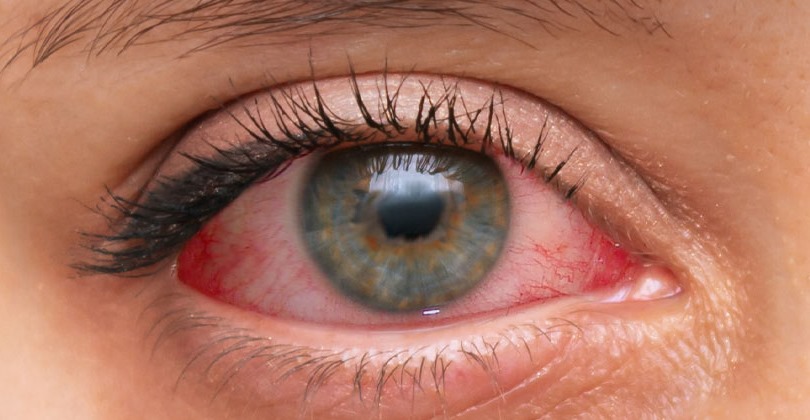
Conjunctivitis usually causes diffuse injection that involves the fornices and palpebral conjunctiva.
However, severe iritis can redden the whole eye, so pattern alone cannot settle the diagnosis.
The Pupil in Iritis vs Conjunctivitis
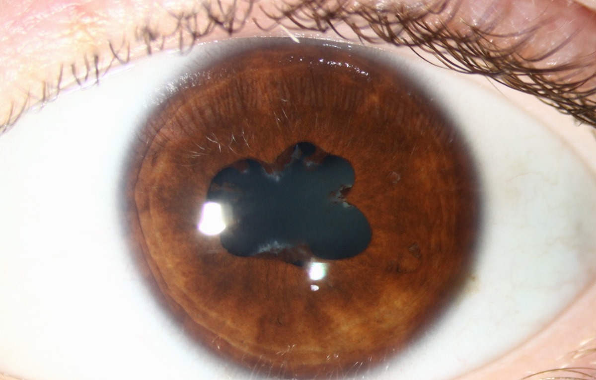
Sphincter spasm often makes the pupil smaller in iritis, and its reaction may slow.
Posterior synechiae can then distort its shape, especially during dilation.
Conjunctivitis leaves the pupil normal.
Meanwhile, a mid-dilated pupil with corneal edema should raise concern for acute angle closure.
Discharge and Tearing
Iritis may cause watering, but it does not usually produce mucopurulent discharge.
Conjunctivitis, however, commonly causes watery, mucoid or purulent discharge.
Copious, rapidly recurring pus suggests gonococcal conjunctivitis, which needs urgent treatment because corneal perforation can occur.
Slit Lamp Signs
Anterior chamber cells define active iritis, and flare reflects leakage of protein into the aqueous.
The SUN system grades both findings, which makes follow-up more consistent.
Also inspect the endothelium, because keratic precipitate patterns can guide the differential.
In conjunctivitis, evert the lids and classify the reaction as follicular or papillary.
Follicles suggest viral or chlamydial disease, while papillae occur in bacterial and allergic disease.
Intraocular Pressure in Iritis vs Conjunctivitis
Pressure usually stays normal in uncomplicated conjunctivitis.
Iritis, however, can lower pressure through reduced aqueous production.
It can also raise pressure, particularly in herpetic disease or with trabecular inflammation.
History That Shifts the Diagnosis
Ask about inflammatory back pain, psoriasis, bowel disease and previous attacks when iritis seems likely.
For conjunctivitis, ask about sick contacts and allergen exposure.
Also ask about contact lens wear, recent eye surgery, trauma and herpes zoster, because these can reveal keratitis or endophthalmitis.
Other Causes of a Red Eye
Episcleritis and scleritis can also mimic both conditions.
However, episcleritis typically blanches with topical phenylephrine, while scleritis causes severe, boring pain.
In addition, keratitis must be excluded, because a corneal infiltrate changes management at once.
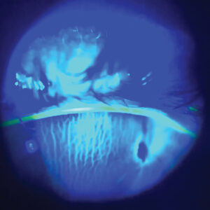
Therefore, stain the cornea with fluorescein in every painful red eye.
Treating Both Conditions
Topical corticosteroids reduce intraocular inflammation in iritis, while cycloplegics relieve spasm and help prevent synechiae.
However, steroids do not treat infection, and they may worsen herpetic disease.
They can also raise pressure and accelerate cataract, so monitoring is essential.
Viral conjunctivitis is usually self-limited, so care focuses on comfort, hygiene and counseling about contagion.
Antibiotics do not shorten viral disease, although they can modestly benefit selected bacterial cases.
Counseling and Follow-Up
First, tell patients with iritis that recurrence is possible and that new symptoms need prompt review.
Next, schedule reassessment within a few days to check cells, pressure and the pupil.
Then taper steroids slowly according to the anterior chamber cell count.
Also advise patients with viral conjunctivitis to wash their hands and avoid sharing towels, because the infection spreads easily.
Referral Triggers in Iritis vs Conjunctivitis
Arrange same-day ophthalmic assessment for any red eye with these features:
- Reduced vision that does not clear with blinking
- Deep pain or marked photophobia
- Ciliary flush or a small, irregular pupil
- Corneal opacity, epithelial defect or hypopyon
- Elevated pressure or copious purulent discharge
- Contact lens wear with pain, recent surgery or trauma
Pitfalls to Avoid
- Do not prescribe steroids for a red eye without a slit lamp examination
- Never assume every red eye with tearing is viral conjunctivitis
- Always evert the lids in chronic conjunctivitis
- Avoid stopping iritis treatment abruptly once the eye looks white


Document what you see
Two smartphone imaging tools built for everyday clinic use — one for the slit lamp, one for the fundus.
From Choroida — the team behind this siteReferences
- Azari AA, Barney NP. Conjunctivitis: a systematic review of diagnosis and treatment. JAMA. 2013;310:1721-1729.
- Jabs DA, Nussenblatt RB, Rosenbaum JT; SUN Working Group. Standardization of uveitis nomenclature for reporting clinical data. Am J Ophthalmol. 2005;140:509-516.
- Cronau H, Kankanala RR, Mauger T. Diagnosis and management of red eye in primary care. Am Fam Physician. 2010;81:137-144.
- Chang JH, McCluskey PJ, Wakefield D. Acute anterior uveitis and HLA-B27. Surv Ophthalmol. 2005;50:364-388.
- Au YK, Henkind P. Pain elicited by consensual pupillary reflex: a diagnostic test for acute iritis. Lancet. 1981;2:1254-1255.

Writes and reviews the clinical articles on Educate. Every post is checked against peer-reviewed sources before it goes live.