
Case Study
A 47-year-old male presented to an ophthalmology clinic with complaints of occasional blurring of vision in his right eye.
The patient, a smoker with a history of hypertension, reported that these episodes of blurred vision were transient and often occurred during physical exertion or after prolonged periods of reading.
Examination Results:
- Visual Acuity: 20/25 in the right eye and 20/20 in the left eye.
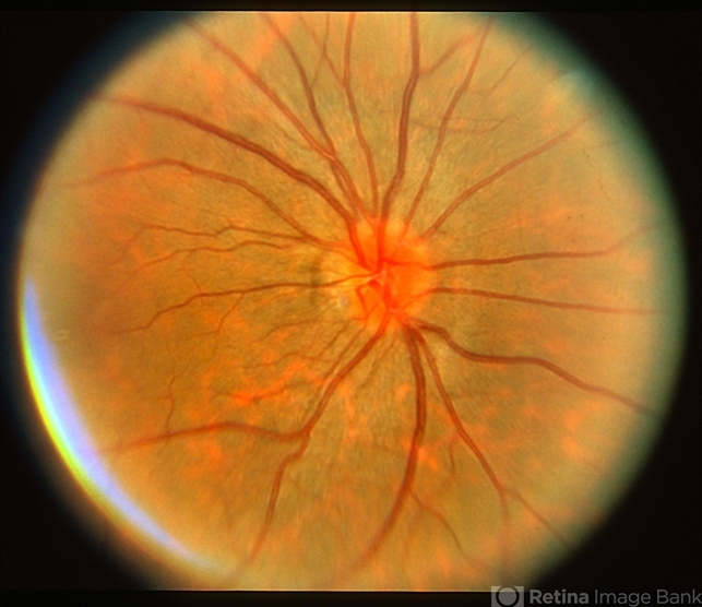
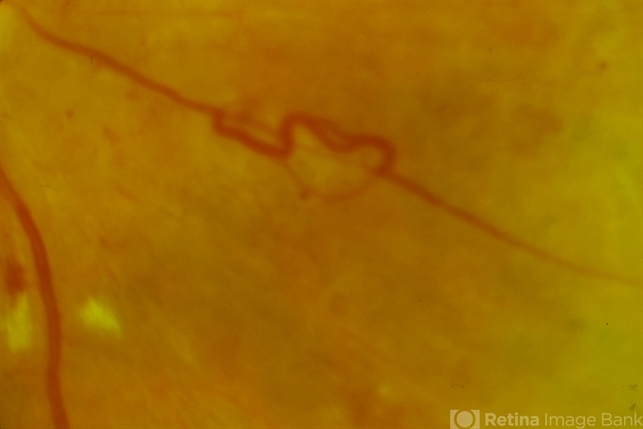
- Fundus Examination: The right eye showed a small, tortuous venous loop located in the mid-peripheral retina, without any associated retinal hemorrhages or exudates.
- Fluorescein Angiography: Revealed a venous loop with normal filling and no evidence of retinal ischemia or capillary nonperfusion.
- Optical Coherence Tomography (OCT): Demonstrated normal macular thickness and no signs of macular edema or structural abnormalities.
- Intraocular Pressure (IOP): 16 mmHg in both eyes.
A diagnosis of a benign Venous Loop was made, and the patient was advised on regular monitoring.
Venous Loop Disease Entity
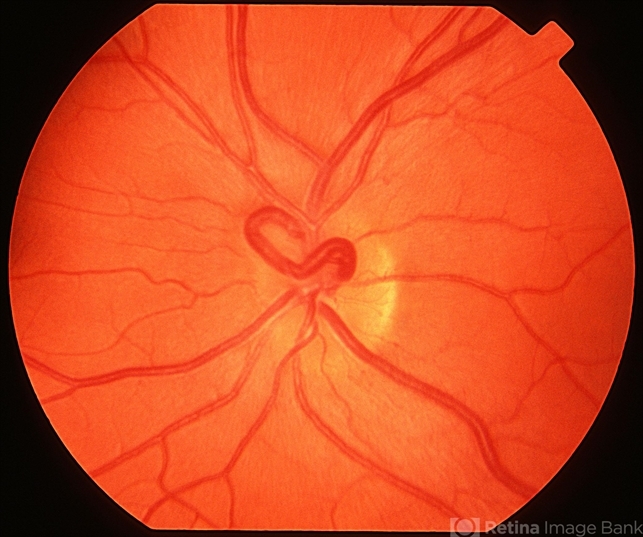
A venous loop is a vascular anomaly characterized by the abnormal tortuosity and looping of a retinal vein.
Venous loops can occur as congenital malformations or as acquired conditions, often associated with systemic vascular diseases.
These loops are typically benign and asymptomatic, but they may occasionally be associated with retinal vein occlusion or other vascular complications.
Venous loops are usually detected incidentally during routine fundus examinations and may appear as a single loop or multiple loops in the retinal periphery.
While most venous loops do not cause significant visual problems, their presence may indicate an underlying vascular pathology, particularly in older patients or those with systemic risk factors such as hypertension or diabetes.
Fundus Explorer Pro
Photograph the retinal findings described here with the phone already in your pocket — 22 D optics and built-in illumination in one handheld unit.
From Choroida — the team behind this sitePathophysiology
The exact pathophysiology of venous loops is not fully understood, but they are thought to result from a combination of genetic predisposition and local hemodynamic factors.
Congenital venous loops likely arise from developmental anomalies during the formation of the retinal vasculature, leading to aberrant connections between retinal veins.
Acquired venous loops may develop in response to chronic vascular stress, such as hypertension or diabetes, which can cause changes in the vascular wall structure and lead to tortuosity.
The localized increase in venous pressure may cause the vessel to loop or twist, creating the characteristic appearance of a venous loop.
While most venous loops remain stable and do not lead to complications, they may predispose the affected area of the retina to ischemia or contribute to the development of retinal vein occlusion, particularly if the loop is associated with other vascular abnormalities.
Venous Loop Epidemiology
Venous loops are relatively rare and are often discovered incidentally during routine eye exams. They can occur at any age but are more commonly observed in older adults, especially those with systemic vascular diseases such as hypertension, diabetes, or atherosclerosis.
The prevalence of venous loops is not well documented, as many cases go unnoticed due to the asymptomatic nature of the condition.
However, they are more likely to be found in individuals with predisposing factors for retinal vascular disease, such as smokers or those with a history of cardiovascular disease.
Clinical Features
Patients with venous loops are often asymptomatic, and the condition is usually detected during routine fundus examination.
However, some patients may present with mild visual symptoms, especially if the loop is associated with other retinal vascular abnormalities:
- Blurred Vision: Transient episodes of blurred vision may occur, particularly if the loop causes localized changes in retinal perfusion.
- Visual Field Defects: Rarely, a venous loop may cause localized visual field defects if it is associated with retinal ischemia.
- No Associated Symptoms: In most cases, venous loops do not cause any symptoms and are discovered incidentally.
Venous Loop Examination Findings
- Fundus Examination: Venous loops appear as small, tortuous loops of retinal veins, typically located in the mid-periphery of the retina. They may be solitary or multiple and are usually not associated with retinal hemorrhages or exudates.
- Fluorescein Angiography: This imaging technique can help to assess the filling pattern of the loop and rule out any associated ischemia or capillary nonperfusion. Most venous loops show normal filling without any signs of leakage.
- Optical Coherence Tomography (OCT): OCT is typically normal in patients with venous loops, as the loops are usually not associated with macular edema or structural retinal changes.
- Intraocular Pressure (IOP): IOP is generally within normal limits in patients with venous loops, but regular monitoring may be necessary to detect any secondary glaucoma.
Differential Diagnosis
The differential diagnosis of venous loops includes other retinal vascular anomalies and conditions that may present with similar fundus findings:
- Retinal Vein Occlusion (RVO): Unlike venous loops, RVO is characterized by sudden vision loss, retinal hemorrhages, and macular edema. Venous loops can, however, predispose to RVO.
- Retinal Microaneurysms: These are small, round outpouchings of retinal capillaries, often seen in diabetic retinopathy, and can sometimes be confused with venous loops.
- Arteriovenous Malformations (AVMs): These are abnormal connections between arteries and veins that can cause retinal hemorrhages and other complications, unlike the usually benign venous loops.
- Vascular Tortuosity: Generalized tortuosity of the retinal vessels may be seen in conditions such as hypertensive retinopathy, but this differs from the localized nature of a venous loop.
Venous Loop Diagnosis
Diagnosis of a venous loop is primarily based on clinical examination and imaging:
- Fundus Examination: The characteristic appearance of a venous loop is usually sufficient for diagnosis.
- Fluorescein Angiography: This can be used to confirm the diagnosis and assess for any associated ischemia or leakage.
- Systemic Workup: Evaluation for underlying systemic vascular conditions may be necessary, particularly in patients with risk factors such as hypertension or diabetes.
Management ofVenous Loop
Management of venous loops generally involves observation and regular monitoring, as most cases do not require intervention:
- Observation: Regular follow-up with fundus examinations and imaging is recommended to monitor for any changes in the loop or development of complications.
- Management of Systemic Risk Factors: Controlling systemic conditions such as hypertension and diabetes is essential to reduce the risk of retinal vascular complications.
- Laser Photocoagulation: In rare cases where a venous loop is associated with retinal ischemia or neovascularization, laser photocoagulation may be considered to prevent further complications.

Prognosis
The prognosis for patients with venous loops is generally excellent, as most loops remain stable and do not lead to significant visual impairment.
However, regular monitoring is essential to detect any potential complications, such as retinal vein occlusion or ischemia, which may affect the visual outcome.
Prevention
Prevention of venous loops primarily involves managing systemic risk factors that may contribute to their development:
- Control of Hypertension and Diabetes: Effective management of these conditions can reduce the risk of developing retinal vascular anomalies, including venous loops.
- Regular Eye Examinations: Routine eye exams are important for early detection of venous loops and other retinal conditions, particularly in individuals with known systemic risk factors.


Document what you see
Two smartphone imaging tools built for everyday clinic use — one for the slit lamp, one for the fundus.
From Choroida — the team behind this siteReferences
- Hayreh, S. S. (1996). Pathogenesis of retinal venous occlusions. Progress in Retinal and Eye Research, 15(4), 507-531.
- McIntosh, R. L., Rogers, S. L., Lim, L., Cheung, N., Wang, J. J., Mitchell, P., & Wong, T. Y. (2010). Natural history of central retinal vein occlusion: an evidence-based systematic review. Ophthalmology, 117(6), 1113-1123.
- McLeod, D. (1997). Retinal vein occlusion: From pathogenesis to treatment. Retina, 17(3), 313-316.
- Klein, R., Klein, B. E., & Moss, S. E. (1996). The epidemiology of retinal vein occlusion: The Beaver Dam Eye Study. Transactions of the American Ophthalmological Society, 94, 593.
- Wooten, B. R., & O’Malley, R. E. (2001). Evaluation and management of retinal venous loops: A case study and review of the literature.
