
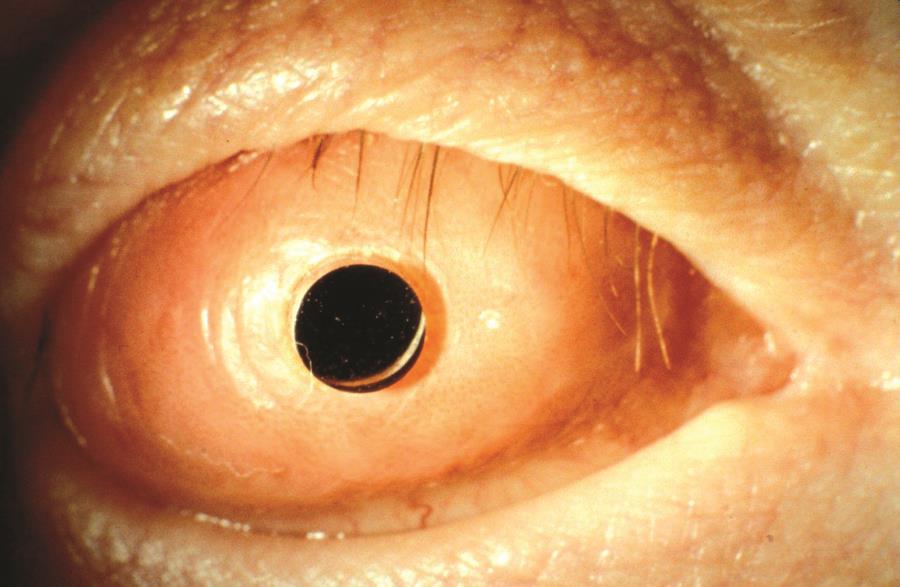
The osteo-odonto-keratoprosthesis, often abbreviated OOKP, is one of the most unusual and biologically ingenious procedures in ophthalmic surgery, using a patient’s own tooth root and surrounding bone as a living scaffold to support an optical cylinder, reserved for the most severe cases of corneal blindness where the ocular surface is too hostile for any other form of corneal or keratoprosthesis surgery to survive.
Why This Extreme Approach Exists
Standard corneal transplantation and even more modern keratoprosthesis devices generally require some baseline ocular surface health, adequate tear production, and reasonable eyelid function to succeed.
In the most severe cases of ocular surface failure, such as end-stage Stevens-Johnson syndrome, severe chemical burns, or advanced ocular cicatricial pemphigoid, the ocular surface is so severely scarred, dry, and inhospitable that conventional grafts and even other keratoprosthesis devices, including the Boston keratoprosthesis, have unacceptably high failure rates, which is the clinical gap the osteo-odonto-keratoprosthesis was developed to fill.
Why a Tooth and Bone
The dentin and alveolar bone surrounding a patient’s own tooth root provide a biologically compatible, well-vascularized, and durable scaffold that the patient’s own body will not reject, since it is autologous tissue, and this composite of tooth root and bone has proven able to survive and support an embedded optical cylinder for years to decades in the harsh, poorly lubricated environment where other materials would be extruded or fail.
All-fit Slit-Lamp Adapter
Record and share exactly what you see at the slit lamp. One adapter fits any slit lamp or surgical microscope — and any smartphone.
From Choroida — the team behind this siteSurgical Procedure
The osteo-odonto-keratoprosthesis is performed in stages, typically over several months.
Stage 1
- A single-rooted tooth, usually a canine, along with a block of surrounding alveolar bone, is harvested from the patient
- The tooth-bone complex (the “lamina”) is shaped and an optical cylinder is embedded within it
- The prepared lamina is then implanted temporarily beneath the skin, typically in the cheek or periorbital region, allowing it to develop its own blood supply (vascularization) over the following months
- Simultaneously, the severely scarred ocular surface is prepared, often with a buccal mucous membrane graft to create a healthier surface for the second stage
Stage 2
- Once the lamina has developed adequate vascularization, it is surgically removed from its temporary site and implanted into the eye
- The optical cylinder is positioned to project through a central opening, providing a clear optical pathway to the retina, while the surrounding vascularized bone-tooth complex, covered by the earlier mucous membrane graft, provides a durable, biologically integrated housing
Indications
The osteo-odonto-keratoprosthesis is reserved for the most severe, bilateral cases of corneal blindness with a severely compromised ocular surface unsuitable for other approaches, including:
- End-stage Stevens-Johnson syndrome and toxic epidermal necrolysis with severe ocular surface scarring
- Severe chemical or thermal ocular burns with extensive limbal stem cell deficiency and surface scarring
- Advanced ocular cicatricial pemphigoid
- Other causes of severe bilateral limbal stem cell deficiency where standard limbal stem cell transplantation has failed or is not expected to succeed given the hostile surface environment
Outcomes and Considerations
Long-term studies have demonstrated impressive anatomic survival of the tooth-bone lamina in a substantial majority of patients over many years, offering a chance at useful vision in patients who would otherwise have essentially no other surgical option.
- The procedure is technically demanding, resource-intensive, and available at only a limited number of specialized centers worldwide
- Ongoing lifelong follow-up is needed, including monitoring for glaucoma, which is common in this patient population, and for resorption of the bone-tooth lamina over very long-term follow-up in some patients
- Patients require realistic counseling about the complexity, staged nature, and long recovery period involved
Alternatives
For patients with severe ocular surface disease not quite severe enough to require an OOKP, or in centers without OOKP expertise, other keratoprosthesis options, including the Boston keratoprosthesis type II (designed specifically for severe surface disease), are considered, though outcomes in the most hostile ocular surfaces generally favor the OOKP given its unique biological durability (see Boston keratoprosthesis).
Prognosis
For appropriately selected patients with the most severe forms of bilateral corneal blindness and ocular surface failure, the osteo-odonto-keratoprosthesis offers a meaningful chance of restoring useful vision where essentially no other option exists, representing one of the more remarkable examples of the body’s own tissue being repurposed to restore sight.


Document what you see
Two smartphone imaging tools built for everyday clinic use — one for the slit lamp, one for the fundus.
From Choroida — the team behind this siteReferences
- Liu C, Okera S, Tandon R, Herold J, Hull C, Thorp S. Visual rehabilitation in end-stage inflammatory ocular surface disease with the osteo-odonto-keratoprosthesis: results from the UK. Br J Ophthalmol. 2008;92:1211-1217.
- Tan A, Tan DT, Tan XW, Mehta JS. Osteo-odonto keratoprosthesis: systematic review of surgical outcomes and complication rates. Ocul Surf. 2012;10:15-25.
- Falcinelli G, Falsini B, Taloni M, Colliardo P, Falcinelli G. Modified osteo-odonto-keratoprosthesis for treatment of corneal blindness: long-term anatomical and functional outcomes. Arch Ophthalmol. 2005;123:1319-1329.
- Hille K, Grabner G, Liu C, et al. Standards for modified osteoodontokeratoprosthesis (OOKP) surgery. Cornea. 2005;24:895-908.
