
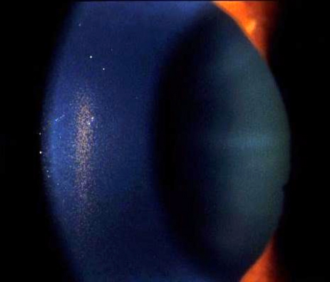
A Krukenberg spindle is a vertically oriented, spindle-shaped band of pigment deposited on the central corneal endothelium, and its characteristic shape and orientation are not random, they directly reflect the normal convection currents of the aqueous humor, giving this classic finding a genuinely elegant mechanistic explanation rather than being simply an arbitrary pattern.
Why the Pigment Forms a Vertical Spindle
In pigment dispersion syndrome, pigment released from the posterior iris pigment epithelium, through chronic mechanical contact with the anterior zonular fibers, circulates within the aqueous humor and settles onto the corneal endothelium following the eye’s normal aqueous convection currents, which move in a pattern influenced by the temperature gradient between the relatively warmer central cornea and cooler periphery.
This convection pattern concentrates settling pigment centrally and in a vertically elongated distribution, producing the characteristic spindle shape, typically widest at its mid-portion and tapering toward the superior and inferior poles of the deposit.
Clinical Appearance
- A vertically oriented, brown to golden-brown, spindle or teardrop-shaped pigment deposit on the central corneal endothelium
- Best visualized with specular reflection or broad, oblique slit-lamp illumination, which enhances the contrast of the pigment deposit against the surrounding endothelial surface
- Size and density can vary considerably between affected individuals, and the spindle can be subtle in earlier or milder disease
- Generally bilateral, though asymmetry between the two eyes is common
All-fit Slit-Lamp Adapter
Record and share exactly what you see at the slit lamp. One adapter fits any slit lamp or surgical microscope — and any smartphone.
From Choroida — the team behind this siteAssociation With Pigment Dispersion Syndrome
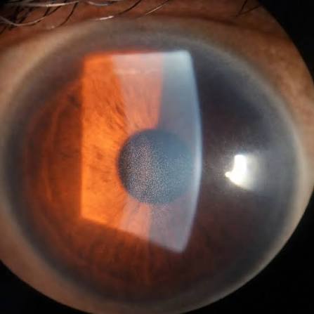
A Krukenberg spindle is one of the classic triad of findings in pigment dispersion syndrome, alongside mid-peripheral iris transillumination defects and increased, often densely homogeneous, trabecular meshwork pigmentation on gonioscopy, and its presence should prompt a complete evaluation for this broader syndrome when it has not already been identified (see trabecular meshwork pigmentation and iris transillumination defects for the related findings in this same triad).
Clinical Significance Beyond the Spindle Itself
The Krukenberg spindle itself does not affect vision or corneal function in any meaningful way, and its clinical significance lies entirely in what it indicates about the broader underlying condition: active pigment dispersion, which carries a real risk of elevated intraocular pressure and pigmentary glaucoma in a meaningful proportion of affected patients.
- Younger, myopic patients are disproportionately affected by pigment dispersion syndrome, and identifying a Krukenberg spindle in this demographic should prompt particular attention to intraocular pressure and optic nerve assessment
- The degree of pigment dispersion activity, reflected in part by the density of the Krukenberg spindle and trabecular pigmentation, does not perfectly predict glaucoma risk, so ongoing monitoring remains important even in patients with a relatively subtle spindle
Diagnostic Evaluation
- Slit-lamp examination with specular or oblique illumination specifically directed at the central corneal endothelium to identify and characterize the spindle
- A complete assessment for the other components of the pigment dispersion triad, including gonioscopy and iris transillumination assessment
- Intraocular pressure measurement and optic nerve evaluation, given the recognized glaucoma risk associated with this underlying syndrome
- Refractive history, given the well-documented association between pigment dispersion syndrome and myopia
Management
The Krukenberg spindle itself requires no direct treatment, since it is an asymptomatic, functionally insignificant corneal finding in its own right.
Management is directed at the broader pigment dispersion syndrome and, when present, any resulting pigmentary glaucoma, following standard glaucoma management principles, including topical aqueous-suppressant medical therapy, with escalation to laser or surgical treatment when needed for inadequately controlled pressure, alongside sustained, ongoing monitoring given the lifelong nature of the underlying pigment-releasing process.
Who gets it
The spindle is classically found in young, myopic patients, often men, with a deep anterior chamber and a posteriorly bowed iris. The vertical band of pigment on the endothelium is formed by the convection currents of aqueous. Patients may notice halos after exercise or dilation, when pigment is released in showers and the pressure spikes.
Management points
Check the pressure at several visits and after exercise if symptoms suggest it. Examine the angle for dense pigment, the iris for transillumination defects, and the peripheral retina for lattice degeneration, which is more common in these myopic eyes. Laser peripheral iridotomy may reduce iris bowing in selected young patients, though evidence for preventing glaucoma is limited. Advise that regular follow-up is needed, because pigmentary glaucoma may develop.
Counselling
Explain that the spindle is a sign of pigment release and not the disease itself, that many people never develop glaucoma, and that pressure checks are the safeguard. Encourage regular examinations and avoidance of unnecessary dilation.
Prognosis
The corneal finding itself carries an excellent prognosis and requires no monitoring beyond its value as a marker of the underlying pigment dispersion process.
The broader prognosis depends on whether and how significantly the associated pigment dispersion syndrome affects intraocular pressure and the optic nerve over time, which is why identifying a Krukenberg spindle, rather than noting it as an isolated curiosity, should reliably prompt the complete glaucoma-focused evaluation this finding is really pointing toward.


Document what you see
Two smartphone imaging tools built for everyday clinic use — one for the slit lamp, one for the fundus.
From Choroida — the team behind this siteReferences
- Campbell DG. Pigmentary dispersion and glaucoma. A new theory. Arch Ophthalmol. 1979;97:1667-1672.
- Ritch R. A unification hypothesis of pigment dispersion syndrome. Trans Am Ophthalmol Soc. 1996;94:381-405.
- Farrar SM, Shields MB. Current concepts in pigmentary glaucoma. Surv Ophthalmol. 1993;37:233-252.
- Niyadurupola N, Broadway DC. Pigment dispersion syndrome and pigmentary glaucoma–a major review. Clin Exp Ophthalmol. 2008;36:868-882.
