
Lattice corneal dystrophy is a bilateral, progressive stromal dystrophy caused by abnormal deposition of amyloid within the corneal stroma, producing the characteristic branching, refractile linear lesions that give the condition its name and that are, in the right clinical context, pathognomonic on slit-lamp exam.
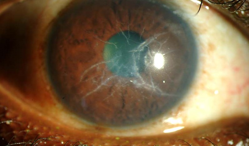
Its particular significance among the corneal stromal dystrophies is that one specific genetic subtype is not confined to the eye at all: it is a marker of a systemic amyloidosis syndrome that can affect nerves, skin, and other organs, making eye exam findings in this specific variant a genuine window into disease well beyond the cornea.
Genetics and Classification
- Lattice type I (classic) — autosomal dominant, caused by mutations in TGFBI, confined to the cornea without systemic amyloid involvement
- Lattice type II (Meretoja syndrome) — caused by mutations in GSN (gelsolin), part of a systemic amyloidosis syndrome that also produces cranial and peripheral neuropathy, characteristic facial skin changes (cutis laxa-like loose, sagging facial skin), and other systemic amyloid deposition; the corneal lattice lines in this variant are typically finer, less dense, and involve a different distribution than classic type I
- Other, rarer variants and combined phenotypes have also been described, generally sharing the underlying theme of localized (or, in type II, systemic) amyloid deposition
The clinical importance of correctly distinguishing type I from type II lattice dystrophy is high: a patient with type II needs systemic evaluation and monitoring for progressive neuropathy and other amyloid-related organ involvement that a patient with isolated type I does not, making the ophthalmologist’s recognition of the specific corneal pattern a potential trigger for a much broader diagnostic workup.
Clinical Presentation
Type I lattice dystrophy typically presents in the first or second decade of life with recurrent corneal erosion episodes — often the earliest symptomatic manifestation, before the lattice lines themselves are dense enough to significantly affect vision — followed by gradual visual decline over subsequent decades as amyloid deposition progressively clouds the central cornea.
Type II (Meretoja syndrome) tends to present somewhat later and progresses more slowly in terms of visual impact, though the systemic neuropathy in this variant can itself be significantly disabling independent of the ocular findings.
All-fit Slit-Lamp Adapter
Record and share exactly what you see at the slit lamp. One adapter fits any slit lamp or surgical microscope — and any smartphone.
From Choroida — the team behind this siteExam Findings
- Refractile, branching, linear opacities (the “lattice” lines) within the corneal stroma, most visible on retroillumination, typically sparing the peripheral cornea and most prominent centrally
- Interspersed white-gray stromal haze between the lattice lines, which progressively increases and is what ultimately drives the visual decline as the disease advances
- Recurrent epithelial erosions, particularly early in the disease course, following the same general principles discussed in this site’s dedicated article on recurrent corneal erosion syndrome
- In type II specifically: additional signs of systemic amyloidosis on general exam — characteristic facial skin laxity, evidence of peripheral or cranial neuropathy (including, notably, facial nerve palsy in some patients), and other findings reflecting the systemic nature of this variant
Differential Diagnosis
- Granular corneal dystrophy — discrete, sharply demarcated, breadcrumb-like deposits with clear intervening stroma, rather than the branching linear pattern of lattice dystrophy, discussed in its own dedicated article on this site
- Macular corneal dystrophy — diffuse, cloudy stromal haze without the discrete linear or granular deposits of the other two classic stromal dystrophies, and notably the only one of the three inherited in an autosomal recessive rather than dominant pattern
- Avellino (granular-lattice) corneal dystrophy — a combined phenotype showing features of both granular and lattice dystrophy, related to specific TGFBI mutation variants
- Amyloid deposition from other, unrelated causes — generally distinguished by the specific clinical and family history context, and confirmed if needed by the same anti-amyloid histochemical staining used to characterize hereditary lattice dystrophy

Management
Recurrent corneal erosion episodes are managed with the same general approach used for that syndrome from any cause: aggressive lubrication as the first line, with more definitive interventions (anterior stromal micropuncture, phototherapeutic keratectomy) reserved for refractory cases, discussed in more detail in this site’s dedicated article on recurrent corneal erosion.
As stromal haze progressively affects vision, phototherapeutic keratectomy can provide temporary improvement by removing more superficial deposits, though the disease is progressive and deposits typically recur over subsequent years, given that the treatment addresses existing amyloid rather than the underlying ongoing deposition process.
Corneal transplantation (penetrating or, increasingly, deep anterior lamellar keratoplasty when the deposition is confined to the stroma without endothelial involvement) is eventually needed in many patients as the disease progresses.
However, a notable and clinically important feature of lattice dystrophy is that amyloid deposition can recur in the graft itself over subsequent years, because the underlying disease process — systemic in type II — is not cured by replacing the affected corneal tissue alone, and patients should be counseled about this possibility before undergoing transplantation.
Patients with type II (Meretoja syndrome) require coordinated care with neurology and other relevant specialists for their systemic amyloid-related manifestations, extending management well beyond the eye alone.
Because the ophthalmologist is sometimes the first clinician to recognize the distinctive lattice pattern, correctly distinguishing type I from type II on exam has real downstream consequences: a type II diagnosis should prompt referral for systemic evaluation rather than being managed purely as a corneal problem.


Document what you see
Two smartphone imaging tools built for everyday clinic use — one for the slit lamp, one for the fundus.
From Choroida — the team behind this siteReferences
- Meretoja J. Familial systemic paramyloidosis with lattice dystrophy of the cornea, progressive cranial neuropathy, skin changes and various internal symptoms. Annals of Clinical Research.
- Klintworth GK. Corneal dystrophies. Orphanet Journal of Rare Diseases.
- American Academy of Ophthalmology. Basic and Clinical Science Course, Section 8: External Disease and Cornea.
Test yourself
A few questions straight from this article.
-
Which material is deposited in the cornea in lattice corneal dystrophy?
Lattice dystrophy is a bilateral progressive stromal dystrophy caused by abnormal amyloid deposition within the corneal stroma, which forms the branching refractile lines. -
Which gene and inheritance pattern underlie classic lattice corneal dystrophy type I?
Classic type I lattice dystrophy is autosomal dominant and caused by TGFBI mutations, and it stays confined to the cornea without systemic amyloid involvement. -
Which gene is mutated in lattice corneal dystrophy type II (Meretoja syndrome)?
Type II lattice dystrophy arises from mutations in GSN (gelsolin) and forms part of a systemic amyloidosis syndrome rather than an isolated corneal disease. -
Which systemic features accompany lattice corneal dystrophy type II?
Meretoja syndrome produces cranial and peripheral neuropathy, including facial nerve palsy in some patients, along with cutis laxa-like loose sagging facial skin and other systemic amyloid deposition. -
What is usually the earliest symptomatic manifestation of lattice corneal dystrophy type I?
Type I typically presents in the first or second decade with recurrent corneal erosions, before the lattice lines are dense enough to affect vision significantly. -
Which finding ultimately drives visual decline in advancing lattice corneal dystrophy?
Interspersed white-gray stromal haze between the lattice lines progressively increases, and it is this haze rather than the lines alone that ultimately reduces vision. -
Which appearance distinguishes granular corneal dystrophy from lattice corneal dystrophy?
Granular dystrophy shows discrete, sharply demarcated breadcrumb-like deposits separated by clear stroma, unlike the branching linear pattern of lattice dystrophy. -
What is first-line treatment for the recurrent erosion episodes of lattice corneal dystrophy?
Recurrent erosions are managed first with aggressive lubrication, with micropuncture or phototherapeutic keratectomy reserved for refractory cases. -
What should patients with lattice corneal dystrophy be counselled about before corneal transplantation?
Amyloid can recur in the graft over subsequent years because replacing the corneal tissue does not cure the underlying deposition process, so patients should be counselled beforehand. -
Recognising lattice corneal dystrophy type II on slit-lamp exam should prompt which action?
A type II diagnosis should trigger referral for systemic evaluation and coordinated care with neurology for progressive neuropathy and other amyloid-related organ involvement, rather than purely corneal management.
