
Retinopathy of Prematurity (ROP) is a potentially blinding eye disorder that primarily affects premature infants.

It is characterized by abnormal development of retinal blood vessels, which can lead to retinal detachment and vision loss if not promptly diagnosed and treated.
Advances in neonatal care have increased the survival rates of preterm infants, but this has also led to a rise in the incidence of Retinopathy of Prematurity (ROP).
Understanding the pathophysiology, risk factors, clinical features, diagnosis, and management of ROP is crucial for preventing and mitigating its impact on vision.
Pathophysiology
ROP occurs in two phases. The first phase begins shortly after birth when the normal growth of retinal blood vessels is interrupted by preterm birth and exposure to extrauterine oxygen.
This leads to a period of vascular arrest and regression. The second phase is marked by pathological neovascularization, driven by hypoxia-induced upregulation of vascular endothelial growth factor (VEGF).
The abnormal blood vessels are fragile and prone to leakage, causing retinal edema and hemorrhage. If left untreated, these vessels can lead to fibrovascular proliferation and tractional retinal detachment.

Retinopathy of Prematurity (ROP) Epidemiology
Retinopathy of Prematurity (ROP) primarily affects premature infants with a birth weight of less than 1500 grams or a gestational age of less than 32 weeks.
The incidence of ROP is inversely proportional to birth weight and gestational age, with the most severe cases occurring in the smallest and most premature infants.
Advances in neonatal care have increased the survival rates of extremely preterm infants, but this has also led to a higher prevalence of ROP in this population.
Fundus Explorer Pro
Photograph the retinal findings described here with the phone already in your pocket — 22 D optics and built-in illumination in one handheld unit.
From Choroida — the team behind this siteRisk Factors
Several factors increase the risk of developing ROP, including:
- Prematurity: The earlier the gestational age at birth, the higher the risk of ROP.
- Low Birth Weight: Infants weighing less than 1500 grams are at significant risk.
- Oxygen Therapy: Prolonged exposure to high levels of supplemental oxygen can disrupt normal retinal vascular development.
- Sepsis: Systemic infections can exacerbate retinal hypoxia and inflammation.
- Blood Transfusions: Frequent transfusions can alter oxygen delivery and contribute to retinal vascular abnormalities.
- Poor Postnatal Growth: Inadequate weight gain after birth is associated with an increased risk of severe ROP.
Clinical Features
Retinopathy of Prematurity (ROP) is classified into five stages based on the severity of retinal changes:
- Stage 1: Mildly abnormal blood vessel growth characterized by a thin demarcation line between vascularized and avascular retina.
- Stage 2: Moderately abnormal blood vessel growth with a ridge of tissue at the junction between the vascularized and avascular retina.
- Stage 3: Severe abnormal blood vessel growth with extraretinal fibrovascular proliferation.
- Stage 4: Partial retinal detachment.
- Stage 5: Total retinal detachment.
Diagnosis
Early diagnosis of Retinopathy of Prematurity (ROP) is essential to prevent progression to severe stages. Screening guidelines recommend that all infants with a birth weight of less than 1500 grams or a gestational age of less than 30 weeks undergo regular retinal examinations starting at 4 to 6 weeks postnatally.
The examination is performed using indirect ophthalmoscopy, often under sedation, to visualize the retina and assess the stage of ROP.

Retinopathy of Prematurity (ROP) Management
The management of ROP depends on the stage and severity of the disease:
- Observation: Infants with mild ROP (Stages 1 and 2 without plus disease) are closely monitored with regular follow-up exams, as many cases spontaneously regress without intervention.
- Laser Photocoagulation: This is the standard treatment for severe ROP (Stage 3 with plus disease). Laser therapy ablates the avascular retina, reducing VEGF production and halting the progression of neovascularization.
- Anti-VEGF Therapy: Intravitreal injections of anti-VEGF agents, such as bevacizumab, are increasingly used for severe ROP. These drugs inhibit VEGF and reduce neovascularization. Anti-VEGF therapy is particularly beneficial for Zone I ROP, which is closer to the optic nerve and more challenging to treat with laser.
- Surgery: In advanced cases (Stages 4 and 5), surgical interventions such as scleral buckling or vitrectomy may be necessary to reattach the retina and preserve vision.
Prognosis
The prognosis for infants with ROP varies depending on the stage at diagnosis and the effectiveness of treatment.
Early stages of ROP often regress spontaneously or respond well to treatment, resulting in favorable visual outcomes.
However, severe ROP can lead to significant visual impairment or blindness despite intervention. Regular follow-up and early intervention are crucial for optimizing visual outcomes in affected infants.
Complications
ROP can lead to several long-term complications, including:
- Myopia: High levels of nearsightedness are common in children with a history of ROP.
- Strabismus: Misalignment of the eyes can occur, requiring corrective measures.
- Amblyopia: “Lazy eye” may develop if one eye is more affected than the other.
- Glaucoma: Increased intraocular pressure can lead to optic nerve damage and vision loss.
- Retinal Detachment: This remains a significant risk in advanced ROP and can result in permanent vision loss.
Role of Smartphone Fundoscopy in ROP
Advances in technology have introduced the potential for smartphone fundoscopy to play a role in the screening and management of Retinopathy of Prematurity (ROP).
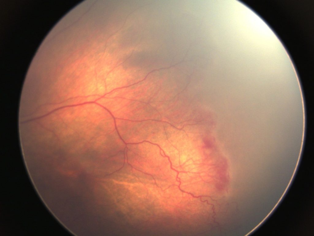
Smartphone-based retinal imaging systems are portable, cost-effective, and can capture high-resolution images of the retina.
These systems allow for remote screening and telemedicine consultations, enabling ophthalmologists to assess retinal images and make timely decisions regarding the need for intervention.
Smartphone fundoscopy (Fundus Explorer Pro) can enhance access to ROP screening in remote or underserved areas, improving early detection and treatment outcomes for premature infants at risk of this blinding condition.


Document what you see
Two smartphone imaging tools built for everyday clinic use — one for the slit lamp, one for the fundus.
From Choroida — the team behind this siteReferences
- Blencowe, H., Lawn, J.E., Vazquez, T., Fielder, A., & Gilbert, C. (2013). Preterm-associated visual impairment and estimates of retinopathy of prematurity at regional and global levels for 2010. Pediatric Research, 74(S1), 35-49.
- Fierson, W.M., Capone, A., & American Academy of Pediatrics Section on Ophthalmology. (2018). Screening examination of premature infants for retinopathy of prematurity. Pediatrics, 142(6), e20183061.
- Quinn, G.E., Ying, G.S., Bell, E.F., et al. (2018). Incidence and early course of retinopathy of prematurity: secondary analysis of the postnatal growth and retinopathy of prematurity (G-ROP) study. JAMA Ophthalmology, 136(12), 1383-1390.
- Chen, J., Stahl, A., Hellstrom, A., & Smith, L.E. (2011). Current understanding of the mechanisms of retinopathy of prematurity (ROP). Seminars in Perinatology, 37(4), 296-310.
- Campbell, J.P., Swan, R., Jonas, K.E., et al. (2017). Implementation and evaluation of a tele-education system for the diagnosis of retinopathy of prematurity. Journal of the American Medical Informatics Association, 24(1), 38-44.
Test yourself
A few questions straight from this article.
-
What drives the second phase of retinopathy of prematurity?
After the first phase of vascular arrest and regression, the second phase of ROP is pathological neovascularisation driven by hypoxia-induced upregulation of vascular endothelial growth factor. -
If left untreated, the fragile new vessels of retinopathy of prematurity progress to which outcome?
The abnormal vessels are fragile and leak, causing retinal oedema and haemorrhage, and untreated they go on to fibrovascular proliferation and tractional retinal detachment. -
Below which birth weight are infants considered at significant risk of retinopathy of prematurity?
ROP primarily affects premature infants weighing under 1500 grams at birth, and that weight is the threshold used both for defining risk and for triggering retinal screening. -
How does the incidence of retinopathy of prematurity relate to birth weight and gestational age?
ROP incidence is inversely proportional to birth weight and gestational age, with the most severe disease in the smallest and most premature infants. -
What is the hallmark retinal finding of stage 1 retinopathy of prematurity?
Stage 1 shows mildly abnormal vessel growth marked by a thin demarcation line; the raised ridge at that junction defines stage 2, and extraretinal fibrovascular proliferation defines stage 3. -
How is the retina examined when screening an infant for retinopathy of prematurity?
Screening examinations use indirect ophthalmoscopy, frequently under sedation, to view the retina and assign the stage of ROP. -
How are infants with stage 1 or stage 2 retinopathy of prematurity without plus disease managed?
Mild ROP at stages 1 and 2 without plus disease is simply monitored with regular review, because many such cases regress spontaneously without any intervention. -
How does laser photocoagulation halt the progression of severe retinopathy of prematurity?
Laser therapy, the standard treatment for stage 3 disease with plus disease, ablates the avascular retina, lowering VEGF production and arresting neovascularisation. -
Anti-VEGF therapy is described as particularly beneficial for which pattern of retinopathy of prematurity?
Intravitreal anti-VEGF agents such as bevacizumab are especially useful in Zone I ROP, which lies closer to the optic nerve and is harder to treat with laser. -
Which refractive complication is common in children with a history of retinopathy of prematurity?
High myopia is a common long-term sequela of ROP, alongside strabismus, amblyopia, glaucoma and the continuing risk of retinal detachment.
