Ocular toxoplasmosis recovery follows a predictable sequence once the active lesion stops expanding.
Retinochoroiditis settles first, vitritis clears later, and a pigmented scar forms over the following months. Final vision, however, depends mostly on where the lesion sits.
How Long Ocular Toxoplasmosis Recovery Takes
In immunocompetent patients, active retinochoroiditis is usually self-limited. Most lesions heal within one to two months, although some stay active longer, and large lesions and older patients often take more time. In contrast, immunocompromised patients may develop progressive lesions that do not heal without treatment.
Healing With and Without Treatment
Small peripheral lesions often resolve on their own, so many clinicians observe them in healthy adults. Antiparasitic therapy aims to limit parasite replication and lesion size.
However, a Cochrane review found no clear evidence that antibiotics improve final visual acuity. Rothova and colleagues also found that classic therapy reduced lesion size without shortening the duration of inflammation.
Fundus Explorer Pro
Photograph the retinal findings described here with the phone already in your pocket — 22 D optics and built-in illumination in one handheld unit.
From Choroida — the team behind this siteEarly Signs That the Lesion Is Settling
The first sign of response is a sharper lesion border. Next, the fluffy white edge becomes less opaque and the surrounding retinal edema fades, and the overlying vitreous haze then begins to thin.
Vitritis Clearance in Ocular Toxoplasmosis Recovery
Vitritis lags behind the retinal lesion, and the haze may persist for weeks after the border has sharpened. As a result, vision may stay blurred even when the retina looks quiet. Dense vitreous cells and debris can take several months to clear completely.
Scar Formation Over Time
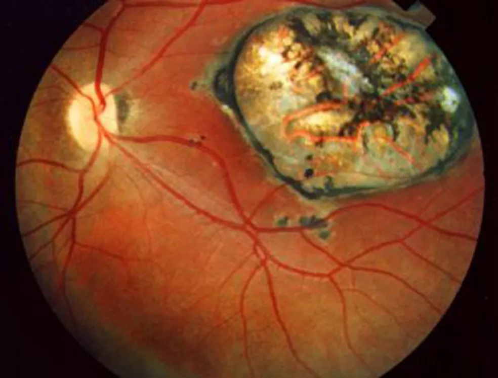
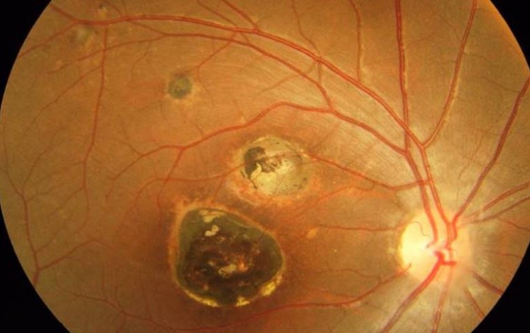
The healed lesion becomes an atrophic chorioretinal scar. Pigment gathers at its border over several weeks, which gives the classic punched-out appearance, and the center often shows bare sclera and visible choroidal vessels. Because the scar harbors encysted parasites, new lesions often appear at its edge (see reading scars for reactivation).
Imaging During Ocular Toxoplasmosis Recovery
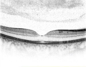
OCT shows a thickened, hyperreflective inner retina during the active phase, and with healing the retina thins and disorganizes while the RPE becomes irregular. Fluorescein angiography also helps when choroidal neovascularization is suspected at the scar margin.
Lesion Location and Ocular Toxoplasmosis Recovery
Location predicts vision better than lesion size. Peripheral scars usually leave good central acuity, while a foveal scar causes a permanent central scotoma. However, perifoveal lesions may reduce acuity through macular edema while active, but that edema usually resolves as the lesion heals, so acuity can improve.
Juxtapapillary Lesions and the Optic Nerve
Lesions next to the disc damage the nerve fiber bundles passing through them, so patients develop a sector or arcuate field defect that often persists. Central acuity may stay normal if the papillomacular bundle is spared. Therefore, document visual fields once the lesion heals.
Complications That Delay Ocular Toxoplasmosis Recovery
Some eyes heal slowly because of secondary problems. For example, choroidal neovascularization can develop at a scar edge months or years later. Epiretinal membrane, cystoid macular edema and vitreous traction may also limit vision, and retinal detachment occasionally follows vitreous contraction.
Role of Corticosteroids
Oral corticosteroids are often added for vision-threatening lesions or severe vitritis, and they aim to reduce inflammatory damage and speed vitreous clearing. However, high-quality trial evidence for this benefit is lacking. Never give steroids without antiparasitic cover, because unopposed steroids can worsen the infection.
Follow-Up Schedule
Review active lesions every one to two weeks until the border sharpens, then lengthen intervals as the vitritis clears. Measure lesion size on photographs and compare with baseline. Continued enlargement despite treatment should prompt a review of the diagnosis and regimen.
Recurrence After Healing
Healing does not mean cure, because tissue cysts remain dormant in the scar. Recurrences are common and tend to cluster in the years after an episode.
Long-term intermittent trimethoprim-sulfamethoxazole reduced recurrences in a randomized trial by Silveira and colleagues. Therefore, prophylaxis may suit patients with frequent recurrences or lesions near the fovea.
Counseling on Ocular Toxoplasmosis Recovery
Explain that floaters may outlast the retinal lesion by weeks and that the scar is permanent, so vision lost from a foveal scar will not return. Patients should report new floaters, blur or pain promptly. Pregnant women and immunocompromised patients need individualized plans (see ocular toxoplasmosis for the workup).
Pitfalls to Avoid
- Do not mistake persistent vitritis for treatment failure when the lesion border is sharp
- Do not promise visual recovery from a foveal scar
- Avoid steroids without antiparasitic cover
- Check fields in juxtapapillary lesions even when acuity is normal
- Remember that new floaters after healing may signal reactivation


Document what you see
Two smartphone imaging tools built for everyday clinic use — one for the slit lamp, one for the fundus.
From Choroida — the team behind this siteReferences
- Holland GN. Ocular toxoplasmosis: a global reassessment. Part II: disease manifestations and management. Am J Ophthalmol. 2004;137:1-17.
- Bosch-Driessen LE, Berendschot TT, Ongkosuwito JV, Rothova A. Ocular toxoplasmosis: clinical features and prognosis of 154 patients. Ophthalmology. 2002;109:869-878.
- Rothova A, Meenken C, Buitenhuis HJ, et al. Therapy for ocular toxoplasmosis. Am J Ophthalmol. 1993;115:517-523.
- Pradhan E, Bhandari S, Gilbert RE, Stanford M. Antibiotics versus no treatment for toxoplasma retinochoroiditis. Cochrane Database Syst Rev. 2016;(5):CD002218.
- Silveira C, Belfort R Jr, Muccioli C, et al. The effect of long-term intermittent trimethoprim/sulfamethoxazole treatment on recurrences of toxoplasmic retinochoroiditis. Am J Ophthalmol. 2002;134:41-46.

Writes and reviews the clinical articles on Educate. Every post is checked against peer-reviewed sources before it goes live.