RP prognosis describes how retinitis pigmentosa progresses over a lifetime, and it varies widely between patients.
Most patients lose peripheral field first and keep central acuity for many years. However, inheritance, genotype and macular complications all shift the timeline.
How RP Prognosis Is Usually Framed
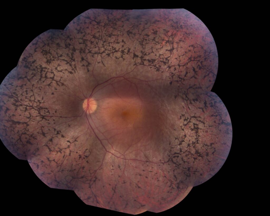
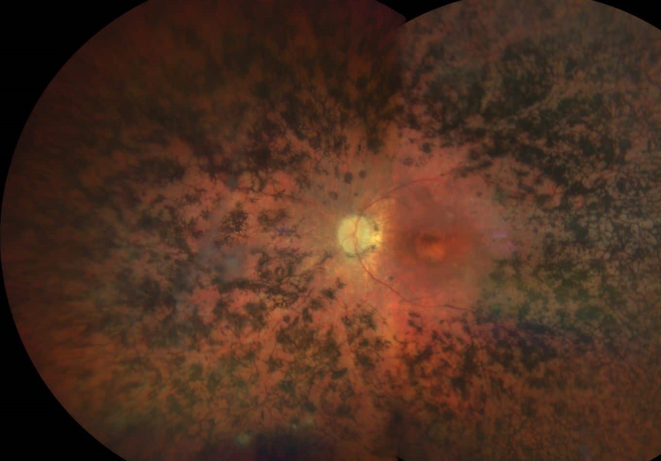
Retinitis pigmentosa is a rod-cone dystrophy, so night vision and midperipheral field fail first, and cone loss follows later while central acuity declines last. Clinicians therefore discuss field survival and central vision survival separately. For the full clinical picture, see our overview of retinitis pigmentosa.
The Natural Rate of Field Loss
Berson and colleagues followed large cohorts with Goldmann fields and full-field ERG, and they found that patients lose a roughly constant fraction of remaining field area each year. As a result, absolute loss is larger early and smaller later.
Fundus Explorer Pro
Photograph the retinal findings described here with the phone already in your pocket — 22 D optics and built-in illumination in one handheld unit.
From Choroida — the team behind this siteRP Prognosis by Inheritance Pattern
Inheritance gives the first broad estimate, and autosomal dominant disease usually starts later and progresses most slowly. Autosomal recessive disease often begins in childhood and follows an intermediate course. X-linked disease is generally the most severe, with myopia and earlier central loss.
X-Linked Disease and RPGR
Affected males with RPGR mutations often report night blindness in early childhood, and natural history data show steady field and acuity loss through young adulthood. Female carriers usually keep useful vision, while some develop significant disease (see X-linked RP and carrier signs), so examine mothers and sisters when the pattern is unclear.
Genotype Refines RP Prognosis
Specific genes can move the estimate in either direction, for example when some RHO mutations cause sectoral or slowly progressive dominant disease. Genetic testing also identifies candidates for trials and for approved RPE65 gene therapy, although variable expression within one family limits gene-based predictions.
Central Vision Preservation
Many patients keep a small central island for decades, and Grover and colleagues found that about half of patients aged 45 or older kept 20/40 or better in one eye. Acuity often stays good until the remaining field becomes very small, and consequently patients may read well while struggling to walk in dim light.
Legal Blindness and Field Size
In the US, a field of 20 degrees or less in the better eye meets the legal blindness definition. As a result, driving and mobility questions arise before reading fails, so refer early for low vision and orientation training.
Imaging Markers in RP Prognosis
OCT shows the ellipsoid zone shrinking toward the fovea as disease advances. Its width correlates with field extent and declines measurably over time, while autofluorescence often shows a hyperautofluorescent ring around the preserved macula.
Cystoid Macular Edema in RP Prognosis
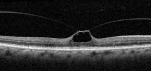
CME affects a meaningful minority of patients and can blur central vision at any stage, and OCT shows intraretinal cysts that often leak little on fluorescein. Carbonic anhydrase inhibitors reduce edema in many eyes, but acuity gains are often modest, and edema may recur (see CME in retinitis pigmentosa). Epiretinal membrane and macular hole can also make one eye fall behind the other.
Cataract and RP Prognosis
Posterior subcapsular cataract develops early and commonly in RP, and even a small opacity matters because the patient relies on a tiny central island. Surgery often improves acuity and glare, although zonular weakness and postoperative CME need attention.
Systemic Clues That Change RP Prognosis
Syndromic forms carry their own outlook, for example Usher syndrome, which adds hearing loss to the retinal disease. Bardet-Biedl syndrome adds obesity, polydactyly and renal disease, and the macula is often involved early, so ask about hearing and kidneys before giving numbers.
Do Supplements Change RP Prognosis?
Berson’s trial suggested vitamin A palmitate slowed cone ERG decline in adults with typical RP. However, a clear benefit on fields and acuity was not shown, and the finding remains debated. High-dose vitamin A also carries liver and teratogenic risks, so discuss it cautiously and avoid it in pregnancy and ABCA4 disease.
Follow-Up Schedule
Annual review suits most stable adults, and each visit should include acuity, fields, OCT and a lens check. Shorten the interval when CME, cataract or rapid change appears, and use the same perimetry strategy each time, because method changes mimic progression.
Counseling on RP Prognosis
Give ranges, and explain that individual eyes vary. First, separate night and side vision from reading vision, and next, explain which complications are treatable. Finally, discuss trials and gene therapy honestly, without promising a cure.
Pitfalls to Avoid
- Do not quote one timeline for all inheritance patterns
- Check for CME when acuity drops faster than expected
- Assess cataract early in a patient with a small central island
- Keep perimetry methods consistent between visits


Document what you see
Two smartphone imaging tools built for everyday clinic use — one for the slit lamp, one for the fundus.
From Choroida — the team behind this siteReferences
- Hartong DT, Berson EL, Dryja TP. Retinitis pigmentosa. Lancet. 2006;368:1795-1809.
- Berson EL, Sandberg MA, Rosner B, et al. Natural course of retinitis pigmentosa over a three-year interval. Am J Ophthalmol. 1985;99:240-251.
- Grover S, Fishman GA, Anderson RJ, et al. Visual acuity impairment in patients with retinitis pigmentosa at age 45 years or older. Ophthalmology. 1999;106:1780-1785.
- Sandberg MA, Rosner B, Weigel-DiFranco C, et al. Disease course of patients with X-linked retinitis pigmentosa due to RPGR gene mutations. Invest Ophthalmol Vis Sci. 2007;48:1298-1304.
- Berson EL, Rosner B, Sandberg MA, et al. A randomized trial of vitamin A and vitamin E supplementation for retinitis pigmentosa. Arch Ophthalmol. 1993;111:761-772.

Writes and reviews the clinical articles on Educate. Every post is checked against peer-reviewed sources before it goes live.