Orbital fat prolapse is a herniation of intraconal orbital fat into the subconjunctival space.
It usually appears as a soft yellow bulge in the superotemporal fornix, and because it looks like a mass, patients and referring clinicians often fear a tumor. However, a short slit-lamp examination usually settles the diagnosis.
What Is Orbital Fat Prolapse
The lesion is normal orbital fat in an abnormal position, and it pushes forward through a weak point in Tenon’s capsule and the intermuscular septum. As a result, it lies directly under the bulbar conjunctiva and carries no malignant potential.
Who Gets Orbital Fat Prolapse
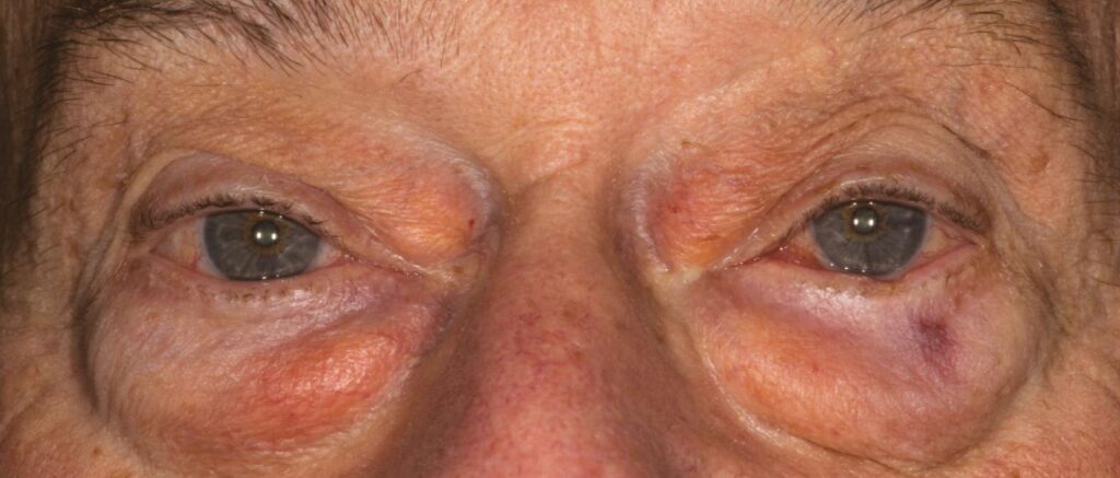
Most patients are older adults, and published series report more men than women. Obesity is a frequent association, and the condition is often bilateral, although one side may look larger.
All-fit Slit-Lamp Adapter
Record and share exactly what you see at the slit lamp. One adapter fits any slit lamp or surgical microscope — and any smartphone.
From Choroida — the team behind this siteWhy the Fat Herniates
Tenon’s capsule thins with age, and then the intraconal fat between the superior and lateral rectus muscles finds a path forward. Raised orbital fat volume, for example in obesity, may add pressure behind the globe, so the bulge typically emerges in the superotemporal quadrant.
Symptoms of Orbital Fat Prolapse
Many patients simply notice the bulge in the mirror, while others describe foreign body sensation, mild irritation or a swelling that shows when they look down. Vision stays normal, and pain, diplopia or rapid growth should therefore prompt a different diagnosis.
Slit-Lamp Appearance
The lesion is yellow, soft, smooth and lobulated, and fine conjunctival vessels run over its surface while the overlying epithelium stays normal. It sits posterior to the limbus and never invades the cornea, and the conjunctiva moves freely over it (see slit-lamp examination technique).
The Reposition Test in Orbital Fat Prolapse
First, ask the patient to look down and in to expose the lesion fully. Next, press gently with a cotton-tipped applicator after topical anesthesia, and prolapsed fat flattens or slides back into the orbit, then bulges again on release. In addition, gentle retropulsion of the globe through the closed lid often enlarges it, so a reducible lesion strongly supports herniated fat.
Orbital Fat Prolapse Versus Dermolipoma
Dermolipoma is a congenital choristoma, so it is usually present from childhood (see dermolipoma). Its surface often looks keratinized and may carry fine hairs, and it is also firmer and adherent to the globe, so it does not reduce with pressure. Orbital fat prolapse, by contrast, appears in adulthood and has a glistening, normal surface.
Orbital Fat Prolapse Versus Conjunctival Lymphoma
Conjunctival lymphoma typically looks salmon-pink and fleshy, but it does not slide back with pressure or change with gaze. Because lymphoma needs biopsy and systemic staging, any pink, fixed lesion deserves tissue (see conjunctival lymphoma).
Other Mimics to Consider
- Prolapsed lacrimal gland: pink, firm and palpable under the superotemporal orbital rim
- Lipoma or liposarcoma: rare, usually firmer, and not reducible
- Conjunctival cyst: translucent, fluid-filled and transilluminates
- Lymphoid hyperplasia: pink and fixed, so it needs biopsy like lymphoma
When to Image
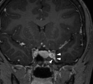
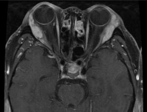
Typical clinical findings usually make imaging unnecessary, but CT or MRI helps when the lesion is atypical, firm or progressive. Imaging then shows fat density continuous with the intraconal fat and excludes a solid mass.
Biopsy and Pathology
Reserve biopsy for lesions that do not behave like fat, and histology shows mature adipocytes, although some specimens also contain atypical stromal or multinucleated cells. For example, Schmack and colleagues showed these features can mimic pleomorphic lipoma or atypical lipomatous tumor, so always tell the pathologist the clinical setting.
Observation in Orbital Fat Prolapse
Most patients need reassurance and nothing more, and lubricating drops can ease irritation from the raised surface. Photograph the lesion at the first visit, then compare its size at follow-up to confirm stability.
Surgical Excision
Consider surgery for cosmetic concern or persistent irritation, in which case the surgeon removes the prolapsed fat through a conjunctival incision and closes the capsular defect. Conservative removal may limit the risk of a hollow appearance, while risks include lateral rectus or levator injury, lacrimal ductule damage with dry eye, and recurrence.
Counseling Patients With Orbital Fat Prolapse
Start by describing the lesion as normal fat in the wrong place, and most patients relax once they see it slide back with the applicator. Next, explain that surgery is optional and carries real risks, and finally, ask them to return if the bulge becomes firm, pink or painful.
Pitfalls to Avoid
- Do not biopsy a soft, reducible yellow bulge in an older adult
- Never label a pink, fixed fornix mass as fat without tissue
- Do not mistake a prolapsed lacrimal gland for fat and excise it


Document what you see
Two smartphone imaging tools built for everyday clinic use — one for the slit lamp, one for the fundus.
From Choroida — the team behind this siteReferences
- Schmack I, Patel RM, Folpe AL, et al. Subconjunctival herniated orbital fat: a benign adipocytic lesion that may mimic pleomorphic lipoma and atypical lipomatous tumor. Am J Surg Pathol. 2007;31:193-198.
- Kim YD, Goldberg RA. Orbital fat prolapse and dermolipoma: two distinct entities. Korean J Ophthalmol. 1994;8:42-43.
- Shields CL, Shields JA. Tumors of the conjunctiva and cornea. Surv Ophthalmol. 2004;49:3-24.
- American Academy of Ophthalmology. Basic and Clinical Science Course, Section 7: Oculofacial Plastic and Orbital Surgery. San Francisco: AAO; 2023.
- Salmon JF. Kanski’s Clinical Ophthalmology: A Systematic Approach. 9th ed. Elsevier; 2020.

Writes and reviews the clinical articles on Educate. Every post is checked against peer-reviewed sources before it goes live.