Anesthetic abuse keratopathy is a toxic corneal disease caused by repeated use of topical anesthetic drops.
Proparacaine and tetracaine numb the eye within seconds, so patients quickly learn to depend on them. However, frequent dosing stops epithelial healing and inflames the stroma, and the result can look like a severe infection.
What Is Anesthetic Abuse Keratopathy
The condition starts with a small epithelial problem that never closes, and then the patient uses anesthetic drops to control the pain. Each drop relieves pain briefly, while it also deepens the corneal injury. As a result, a vicious cycle of pain and toxicity develops.
Why Topical Anesthetics Damage the Cornea
Topical anesthetics inhibit epithelial cell migration and mitosis, and they also disrupt surface microvilli and destabilize the tear film. In addition, they appear toxic to keratocytes and suppress corneal sensation, so the normal protective blink and healing responses fail together.
All-fit Slit-Lamp Adapter
Record and share exactly what you see at the slit lamp. One adapter fits any slit lamp or surgical microscope — and any smartphone.
From Choroida — the team behind this siteWho Develops Anesthetic Abuse Keratopathy
Many patients receive a bottle after a corneal abrasion, welding flash or refractive surgery. Others obtain drops from work, because health care workers can access them easily, and some patients also have depression, anxiety or substance use disorders.
Symptoms Patients Report
Patients describe severe pain, photophobia and blurred vision, and the pain often seems to worsen despite appropriate treatment. However, some patients look surprisingly comfortable during the examination.
Slit Lamp Signs of Anesthetic Abuse Keratopathy
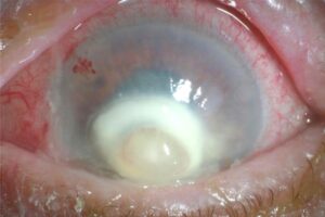
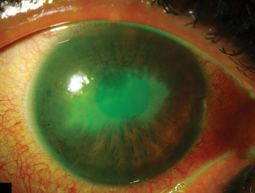
The examiner first sees a persistent epithelial defect with rolled or irregular edges. Next, stromal edema and folds in Descemet membrane often appear, and anterior chamber reaction can be marked, so a sterile hypopyon may form. In advanced cases, the stroma thins and may perforate.
The Ring Infiltrate
A dense stromal ring is the most alarming sign, and it likely reflects immune complex deposition, similar to a Wessely ring. Therefore, its presence does not prove infection (see immune corneal rings), but every ring still demands careful exclusion of microbes.
How Anesthetic Abuse Keratopathy Mimics Acanthamoeba
Both diseases cause severe pain, a ring infiltrate and slow progress, and both also resist standard antibacterial therapy. In addition, routine cultures are often negative in each condition, so clinicians may start long antiamoebic courses without a clear diagnosis (see Acanthamoeba ring infiltrate).
Clues That Separate the Two
Several features help with the distinction:
- Contact lens wear and water exposure favor Acanthamoeba
- Radial perineural infiltrates suggest Acanthamoeba
- Confocal microscopy may show cysts in Acanthamoeba
- Rapid improvement under supervision favors anesthetic toxicity
- A history of eye trauma, welding or recent surgery favors anesthetic abuse
Red Flags in the History
Ask directly whether the patient uses any numbing drops. Many patients deny it at first, so ask again in a neutral way, and finally, repeated visits to emergency departments for eye pain deserve attention.
Laboratory Workup in Anesthetic Abuse Keratopathy
Corneal scraping for Gram stain and cultures remains necessary, and fungal cultures matter because secondary Candida keratitis has been reported. Then consider Acanthamoeba culture or confocal imaging when the history fits. Negative results support the diagnosis, although they never exclude infection completely.
Stopping the Drug
Withdrawal of the anesthetic is the main treatment, but the patient must understand why the drops are causing harm. Some patients need admission so staff can control all medications, and then healing often begins within days of stopping the drug.
Supportive Treatment for Anesthetic Abuse Keratopathy
Use preservative-free lubricants to support the epithelium, and a cycloplegic drop also reduces ciliary spasm and pain. Oral analgesics replace the topical anesthetic during withdrawal, and in addition, a prophylactic antibiotic protects the open defect. A bandage contact lens may help, while close monitoring for infection continues.
Surgical Options
Amniotic membrane transplantation can promote closure of a stubborn defect, and tarsorrhaphy also protects the surface, especially when sensation stays poor. For perforation, tissue adhesive or tectonic keratoplasty may be needed. However, grafts often fail if anesthetic use continues.
Talking With the Patient
Explain the cycle of pain and toxicity in simple words, and avoid blame, because shame often drives hidden use. For example, say that the drops numb the eye while they block healing, and also consider psychiatric referral when dependence or mood disorder is present.
Prognosis of Anesthetic Abuse Keratopathy
Early cessation usually allows the epithelium to heal. Nevertheless, stromal scarring can leave permanent visual loss, and persistent anesthetic use risks perforation and loss of the eye. Therefore, follow patients closely after discharge, because relapse is common, and reduced corneal sensation may persist and overlap with neurotrophic keratopathy.
Pitfalls to Avoid
- Do not escalate antimicrobials endlessly without asking about anesthetic drops
- Never assume a ring infiltrate is always infectious
- Do not perform keratoplasty while the patient still uses the drops
- Avoid confrontation that drives the patient away from follow-up


Document what you see
Two smartphone imaging tools built for everyday clinic use — one for the slit lamp, one for the fundus.
From Choroida — the team behind this siteReferences
- Rosenwasser GO, Holland S, Pflugfelder SC, et al. Topical anesthetic abuse. Ophthalmology. 1990;97:967-972.
- Chern KC, Meisler DM, Wilhelmus KR, et al. Corneal anesthetic abuse and Candida keratitis. Ophthalmology. 1996;103:37-40.
- Yagci A, Bozkurt B, Egrilmez S, et al. Topical anesthetic abuse keratopathy: a commonly overlooked health care problem. Cornea. 2011;30:571-575.
- McGee HT, Fraunfelder FW. Toxicities of topical ophthalmic anesthetics. Expert Opin Drug Saf. 2007;6:637-640.
- Kim JY, Choi YS, Lee JH. Keratitis from corneal anesthetic abuse after photorefractive keratectomy. J Cataract Refract Surg. 1997;23:447-449.

Writes and reviews the clinical articles on Educate. Every post is checked against peer-reviewed sources before it goes live.