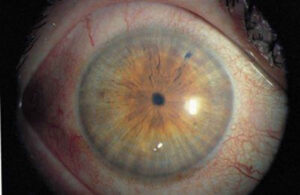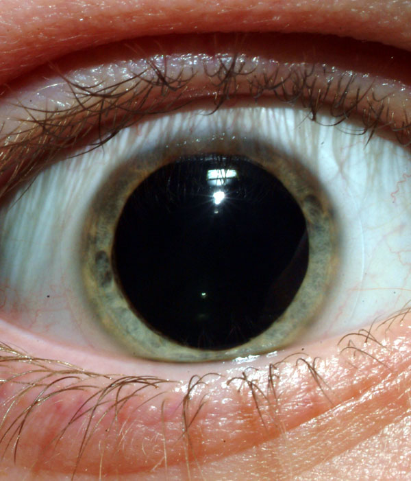
Blown pupil is the bedside term for a unilateral fixed dilated pupil with poor or absent light reaction.
Most causes are benign.
However, a compressive third nerve palsy can be fatal if missed.
Therefore, every examiner needs a fast, structured approach.
What a Blown Pupil Means
Parasympathetic fibers reach the iris sphincter with the third cranial nerve.
Damage along this pathway, or to the sphincter itself, leaves the pupil large and sluggish.
Also, muscarinic blocking drugs produce the same appearance.
First Question: Is the Patient Alert?
A drowsy patient with a new dilated pupil may have uncal herniation.
Therefore, arrange emergency neurosurgical assessment before any detailed eye examination.
The ophthalmic workup below applies to the alert, stable patient.
All-fit Slit-Lamp Adapter
Record and share exactly what you see at the slit lamp. One adapter fits any slit lamp or surgical microscope — and any smartphone.
From Choroida — the team behind this siteConfirm Which Pupil Is Abnormal
First, measure anisocoria in bright and dim light.
If the difference grows in bright light, the larger pupil is abnormal.
Then check the near response and look for segmental sphincter movement at the slit lamp.
Compressive Third Nerve Palsy and the Blown Pupil
A posterior communicating artery aneurysm can compress the superficial pupillomotor fibers.
Because these fibers run on the outer surface of the nerve, the pupil often dilates early.
Look for ptosis, limited adduction, elevation and depression, and new headache.
Treat a painful pupil-involving palsy as an aneurysm until imaging excludes it (see third nerve palsy).
Isolated Blown Pupil Without Ptosis
A fixed dilated pupil with full motility and normal lids rarely comes from an aneurysm.
However, test motility carefully for subtle elevation and adduction deficits.
Also, repeat the examination within hours if the onset was acute.
Pharmacologic Causes
Accidental exposure explains many isolated cases.
For example, ask about scopolamine patches, ipratropium nebulizers, atropine drops, gardening and plants.
In addition, adrenergic agents such as phenylephrine dilate the pupil and blanch the conjunctiva.
These pupils are often very large and show no segmental movement (see pharmacologic mydriasis).
Pilocarpine Testing in a Blown Pupil
Instill 1% pilocarpine in both eyes and recheck after about 30 to 45 minutes.
A pupil that fails to constrict suggests muscarinic blockade or sphincter damage.
By contrast, a third nerve pupil constricts because sphincter receptors remain intact.
Dilute pilocarpine (0.1% or 0.125%) constricts a tonic pupil through denervation supersensitivity.
Traumatic Mydriasis
Blunt injury can tear the sphincter and leave a fixed or irregular pupil.
At the slit lamp, look for margin notches, transillumination defects and hyphema.
Recovery varies, and some pupils improve over weeks, while others stay dilated permanently (see traumatic mydriasis).
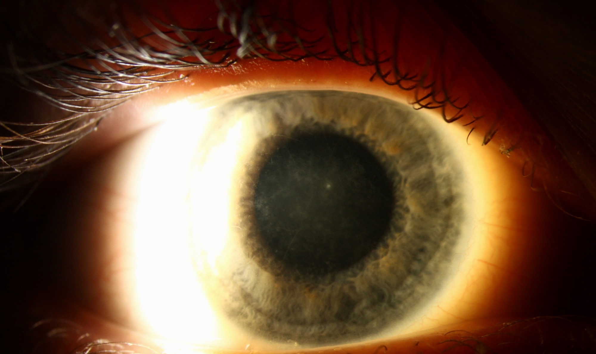
Blown Pupil After Head Trauma
First, exclude intracranial hemorrhage and herniation with urgent CT.
Next, test for a relative afferent pupillary defect, which suggests traumatic optic neuropathy.
However, an afferent defect alone never causes anisocoria.
Adie Tonic Pupil
A tonic pupil usually affects healthy young adults, more often women.
It reacts poorly to light, yet constricts slowly to near and redilates slowly.
Also, the slit lamp shows sectoral palsy with vermiform movements.
Acute Angle Closure Mimicking a Blown Pupil
A mid-dilated, poorly reactive pupil in a red, painful eye suggests angle closure.
Also, the cornea looks hazy, the chamber shallow, and the pressure markedly raised.
Therefore, measure intraocular pressure in every red eye with a large pupil.
Imaging Decisions
A pupil-involving third nerve palsy needs same-day CT angiography or MR angiography.
If results are negative and suspicion stays high, discuss catheter angiography with neurointerventional colleagues.
In contrast, a confirmed pharmacologic pupil needs no imaging.
Triage Checklist for a Blown Pupil
- Drowsiness or new neurological deficit: emergency department now
- Ptosis, diplopia or motility deficit: same-day vascular imaging
- Severe headache with any third nerve sign: treat as aneurysm
- Red painful eye with high pressure: manage as angle closure
- Clear trauma with sphincter tears: slit lamp and pressure check
- No response to 1% pilocarpine: pharmacologic block or iris damage
- Light near dissociation and dilute pilocarpine response: tonic pupil
Counseling the Patient With a Blown Pupil
Explain the cause in plain language once you exclude dangerous causes.
For drug exposure, the pupil usually recovers within days, although atropine can last longer.
In addition, tinted lenses or reading glasses help patients with glare or near blur.
Finally, ask patients to return at once if ptosis, diplopia or headache appear.
Pitfalls to Avoid
- Do not attribute a painful dilated pupil to diabetes
- Never interpret pilocarpine testing before checking motility and lids
- Always test both eyes because the fellow pupil serves as control
- Do not dilate a drowsy patient for fundoscopy


Document what you see
Two smartphone imaging tools built for everyday clinic use — one for the slit lamp, one for the fundus.
From Choroida — the team behind this siteReferences
- Thompson HS, Newsome DA, Loewenfeld IE. The fixed dilated pupil: sudden iridoplegia or mydriatic drops? A simple diagnostic test. Arch Ophthalmol. 1971;86:21-27.
- Thompson HS, Pilley SF. Unequal pupils: a flow chart for sorting out the anisocorias. Surv Ophthalmol. 1976;21:45-48.
- Lee AG, Hayman LA, Brazis PW. The evaluation of isolated third nerve palsy revisited: an update on the evolving role of magnetic resonance, computed tomography, and catheter angiography. Surv Ophthalmol. 2002;47:137-157.
- American Academy of Ophthalmology. Basic and Clinical Science Course, Section 5: Neuro-Ophthalmology. San Francisco: AAO; 2023.
- Bowling B. Kanski’s Clinical Ophthalmology: A Systematic Approach. 8th ed. Elsevier; 2016.

Writes and reviews the clinical articles on Educate. Every post is checked against peer-reviewed sources before it goes live.