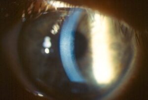
PRK corneal haze is a subepithelial opacity that develops after photorefractive keratectomy as part of the healing response.
Mild haze is common and harmless, but dense haze can reduce vision and may cause regression of the refractive effect.
Modern techniques have lowered its frequency, and the condition remains a topic that every refractive surgeon should know well.
How PRK Corneal Haze Develops
After the surgeon removes the epithelium and ablates the stroma, the cornea heals by activating the keratocytes beneath the ablation.
Some of the keratocytes transform into myofibroblasts, which lay down disorganized collagen and extracellular matrix.
This creates a layer of subepithelial scatter that appears as haze, often several weeks to months after surgery.
Cytokines from the damaged epithelium, including transforming growth factor beta, drive the process, and the epithelial basement membrane plays a role in controlling it.
Time Course
- A clear cornea in the first weeks, with some haze appearing at one to three months
- A peak at around three to six months in most eyes
- Gradual fading over six months to a year in the majority
- A small number with persistent haze, regression of the effect, or both
Patients may notice glare, halos, blurry vision, and increasing myopia as the haze develops.
All-fit Slit-Lamp Adapter
Record and share exactly what you see at the slit lamp. One adapter fits any slit lamp or surgical microscope — and any smartphone.
From Choroida — the team behind this siteRisk Factors of PRK Corneal Haze
- Deeper ablations and higher corrections, which damage more tissue
- Ultraviolet exposure, particularly in the first months
- Atopy, a history of severe dry eye, and some systemic conditions
- Poor epithelial healing or recurrent erosion
- Re-treatment after earlier surface ablation
- Intraoperative factors, such as a large optical zone or rough ablation
Grading
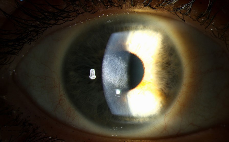
A common scale, by Fantes and colleagues, grades haze from 0 to 4 according to the visibility of iris details.
- Grade 0: a clear cornea
- Trace: a faint haze seen only with oblique illumination
- Grade 1: haze that does not interfere with the view of iris detail
- Grade 2: mild obscuration of iris details
- Grade 3: moderate obscuration of the iris and lens
- Grade 4: a dense opacity that obscures the anterior chamber
Grades 0 to 1 have little effect on vision, whereas grades 2 and above are likely to reduce acuity and cause glare.
Prevention
Intraoperative application of mitomycin C at 0.02 percent for a short time, often 12 to 60 seconds, reduces the risk of haze in higher corrections and in re-treatments.
It works by inhibiting proliferation of keratocytes, and the dose and exposure time are chosen according to the depth of the ablation.
Careful technique, avoidance of excessive ablation depth, and consideration of an alternative procedure for high corrections also help.
Postoperative measures include a taper of topical steroids, sunglasses with ultraviolet protection, and lubricants.
Treatment for PRK Corneal Haze
Mild haze usually needs no treatment and resolves with time.
For moderate haze, topical corticosteroids such as fluorometholone or prednisolone, tapered slowly, may improve the opacity and the refractive result.
Monitor the pressure while on steroids, because some patients are steroid responders (see steroid-induced glaucoma).
Surgeons can treat persistent dense haze by phototherapeutic keratectomy with mitomycin C, which removes the scar and reduces recurrence.
Topical mitomycin C has also been used to treat established haze, although the evidence is limited and the approach has risks, including delayed healing and endothelial toxicity.
Counsel patients about the possible need for further procedures, and about the importance of protecting the eyes from ultraviolet light.
Counselling Before Surgery
Explain that haze is a natural healing response, that it usually fades, and that the chance of a significant problem is small with modern techniques.
Clinicians can offer patients with higher corrections alternatives, including implantable lenses, when appropriate.
PRK Corneal Haze: Differential Diagnosis of a Cloudy Cornea After PRK
Not every opacity after surface ablation is haze, and the surgeon should consider infection, diffuse inflammation, and epithelial healing problems.
A progressive white infiltrate with pain, discharge, and a defect suggests infectious keratitis, which needs culture and immediate treatment.
Persistent epithelial defects, recurrent erosions, and stromal edema can also look like haze.
A careful slit-lamp examination with a narrow beam and attention to the depth of the opacity clarifies the diagnosis (see corneal haze and opacity).
Practical Tips for Follow-Up
Examine patients at one day, one week, one month, and three months after PRK, with a closer schedule for high corrections.
Record the haze grade, the refraction, and the pressure at each visit when steroids are used.
Ask patients about outdoor activity and advise sunglasses with ultraviolet protection for the first year.
If regression of the refractive effect is accompanied by haze, wait until the haze has settled before planning an enhancement.
For the latest studies, see the PubMed results for PRK corneal haze.


Document what you see
Two smartphone imaging tools built for everyday clinic use — one for the slit lamp, one for the fundus.
From Choroida — the team behind this siteReferences
- Netto MV, Mohan RR, Ambrósio R Jr, et al. Wound healing in the cornea: a review of refractive surgery complications and new prospects for therapy. Cornea. 2005;24:509-522.
- Fantes FE, Hanna KD, Waring GO III, et al. Wound healing after excimer laser keratomileusis (photorefractive keratectomy) in monkeys. Arch Ophthalmol. 1990;108:665-675.
- Majmudar PA, Forstot SL, Dennis RF, et al. Topical mitomycin-C for subepithelial fibrosis after refractive corneal surgery. Ophthalmology. 2000;107:2011-2019.
- American Academy of Ophthalmology. Basic and Clinical Science Course, Section 13: Refractive Surgery. AAO; current edition.
Test yourself
A few questions straight from this article.
-
At what corneal level does PRK corneal haze develop after photorefractive keratectomy?
PRK haze is a subepithelial opacity forming as part of the healing response after the stroma is ablated. -
Which cell transformation lays down the disorganized collagen that causes PRK corneal haze?
Keratocytes under the ablation activate and some transform into myofibroblasts, laying down disorganized collagen and matrix that scatter light. -
When does PRK corneal haze typically peak in most eyes?
Haze appears at one to three months, peaks around three to six months, and fades over six months to a year in most eyes. -
The Fantes scale grades PRK corneal haze from 0 to 4 according to what?
The Fantes scale grades haze by how much it obscures the iris, from a clear cornea to a dense opacity obscuring the anterior chamber. -
From which Fantes grade is PRK corneal haze likely to reduce acuity and cause glare?
Grades 0 to 1 have little effect on vision, while grade 2 and above, with obscuration of iris detail, are likely to reduce acuity. -
Which intraoperative measure reduces the risk of PRK corneal haze in higher corrections and re-treatments?
Brief intraoperative mitomycin C 0.02 percent, often 12 to 60 seconds, inhibits keratocyte proliferation and lowers haze risk. -
Moderate PRK corneal haze treated with tapered topical corticosteroids requires monitoring of what?
Fluorometholone or prednisolone, tapered slowly, may improve moderate haze, but pressure must be watched because some patients are steroid responders. -
How is persistent dense PRK corneal haze treated surgically?
Phototherapeutic keratectomy with mitomycin C removes the scar and reduces recurrence in persistent dense haze. -
After PRK, a progressive white infiltrate with pain, discharge and an epithelial defect suggests what instead of haze?
A progressive white infiltrate with pain, discharge and a defect suggests infectious keratitis, which needs culture and immediate treatment. -
When regression after PRK is accompanied by corneal haze, when should an enhancement be planned?
If regression of the refractive effect comes with haze, the surgeon should wait until the haze has settled before planning an enhancement.

Writes and reviews the clinical articles on Educate. Every post is checked against peer-reviewed sources before it goes live.