CRVO vs BRVO compares the two common retinal vein occlusions, which differ in site, extent and risk.
Both cause painless blurring in older adults with vascular risk factors.
However, the fundus pattern, ischemic threat and follow-up schedule differ.
Where Each Occlusion Sits
In CRVO, the thrombus forms at or behind the lamina cribrosa.
As a result, outflow from the whole retina slows.
BRVO usually begins at an arteriovenous crossing, where a thickened artery compresses the vein.
Hypertension, diabetes and hyperlipidemia raise risk in both, while glaucoma links more strongly to CRVO.
Fundus Distribution in CRVO vs BRVO
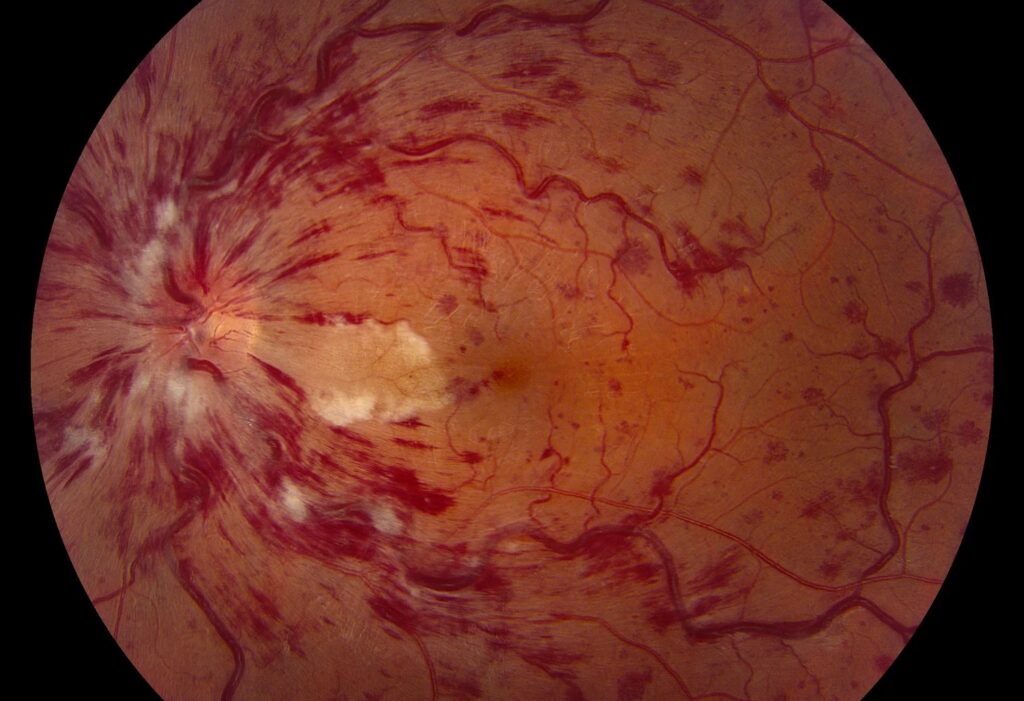
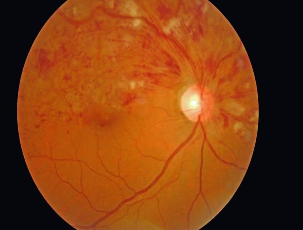
CRVO shows hemorrhages in all four quadrants, with dilated tortuous veins and often disc swelling.
In contrast, BRVO produces a wedge of hemorrhage whose apex points to the crossing.
The superotemporal branch is the commonest site seen in clinic, partly because its occlusion threatens the macula.
Nasal branch occlusions may go unnoticed for months (see arteriovenous crossing changes).
Fundus Explorer Pro
Photograph the retinal findings described here with the phone already in your pocket — 22 D optics and built-in illumination in one handheld unit.
From Choroida — the team behind this siteSymptoms and Presenting Acuity
Patients with CRVO usually describe general blur in one eye.
BRVO causes distortion or a sectoral defect, or no symptoms when the macula is spared.
Because presenting acuity predicts outcome, record it carefully at the first visit.
Ischemia in CRVO vs BRVO
Ischemia drives the most serious complications in both occlusions.
The CVOS defined ischemic CRVO as at least 10 disc areas of nonperfusion on angiography.
A relative afferent pupillary defect, extensive hemorrhage and cotton wool spots also suggest ischemia (see ischemic vs non-ischemic CRVO).
For BRVO, the BVOS used five disc diameters of nonperfusion as its high-risk threshold.
Moreover, about one third of perfused CRVO eyes converted to ischemia within three years in the CVOS.
Macular Edema in CRVO vs BRVO
Macular edema causes most vision loss in both conditions.
CRVO edema usually involves the whole macula and is often severe.
In contrast, BRVO edema tends to be sectoral and may settle as collaterals form.
Because ischemia raises VEGF levels, both forms respond to VEGF blockade.
Anti-VEGF Therapy Evidence
CRUISE showed that ranibizumab improved vision over sham in CRVO edema, and BRAVO showed the same in BRVO.
COPERNICUS and GALILEO then supported aflibercept in CRVO.
In addition, SCORE2 found bevacizumab noninferior to aflibercept for CRVO edema (see anti-VEGF trials in vein occlusion).
Laser Treatment in CRVO vs BRVO
Grid laser plays a different role in each occlusion.
The BVOS showed that grid laser improved vision in BRVO edema with acuity of 20/40 or worse.
However, the CVOS found that grid laser reduced CRVO edema without improving acuity.
Today, laser for BRVO edema mostly serves as an adjunct to injections.
Steroids as Second Line
The SCORE study found triamcinolone better than observation in CRVO edema.
In BRVO, however, it was not superior to grid laser and caused more side effects.
Because cataract and raised pressure follow repeated use, steroids suit pseudophakic or poorly responsive eyes.
Neovascular Risk
Ischemic CRVO carries the highest risk of iris and angle neovascularization.
Neovascular glaucoma often develops within the first months after onset.
BRVO, on the other hand, more often produces disc or retinal new vessels and vitreous hemorrhage.
Scatter Laser Timing in CRVO vs BRVO
The CVOS did not support prophylactic panretinal photocoagulation in ischemic CRVO.
Instead, treat promptly once two clock hours of iris new vessels or any angle vessels appear.
Similarly, the BVOS supported sectoral scatter laser after neovascularization develops in BRVO.
Follow-Up in CRVO vs BRVO
The CVOS examined patients monthly for the first six months.
Many specialists still follow CRVO closely, with undilated iris examination and gonioscopy at each visit.
BRVO usually allows longer intervals once edema is controlled.
Then, injection regimens set the visit frequency in both groups.
Visual Prognosis in CRVO vs BRVO
BRVO generally carries the better prognosis, and many eyes keep useful vision.
CRVO outcomes depend on perfusion status and presenting acuity.
For example, eyes that start with poor acuity seldom regain good vision.
Counseling the Patient
Explain that treatment targets the complications, since no therapy reliably reopens the vein.
Patients with edema should also expect a series of injections over months.
Finally, ask them to report pain, redness or new vision loss promptly.
Pitfalls to Avoid
- Avoid labeling CRVO nonischemic before blood clears enough for angiography
- Never skip gonioscopy in ischemic CRVO
- Check intraocular pressure and the fellow eye
- Exclude diabetic or hypertensive retinopathy before calling bilateral hemorrhages a vein occlusion
- Keep monitoring after the macula dries


Document what you see
Two smartphone imaging tools built for everyday clinic use — one for the slit lamp, one for the fundus.
From Choroida — the team behind this siteReferences
- The Central Vein Occlusion Study Group. Natural history and clinical management of central retinal vein occlusion. Arch Ophthalmol. 1997;115:486-491.
- The Branch Vein Occlusion Study Group. Argon laser photocoagulation for macular edema in branch vein occlusion. Am J Ophthalmol. 1984;98:271-282.
- Campochiaro PA, Heier JS, Feiner L, et al. Ranibizumab for macular edema following branch retinal vein occlusion: six-month primary end point results of a phase III study. Ophthalmology. 2010;117:1102-1112.
- Brown DM, Campochiaro PA, Singh RP, et al. Ranibizumab for macular edema following central retinal vein occlusion: six-month primary end point results of a phase III study. Ophthalmology. 2010;117:1124-1133.
- Scott IU, VanVeldhuisen PC, Ip MS, et al. Effect of bevacizumab vs aflibercept on visual acuity among patients with macular edema due to central retinal vein occlusion: the SCORE2 randomized clinical trial. JAMA. 2017;317:2072-2087.

Writes and reviews the clinical articles on Educate. Every post is checked against peer-reviewed sources before it goes live.