Optic atrophy types describe how a pale optic disc looks and where the original injury probably occurred.
Atrophy is the end stage of axonal loss, so pallor marks damage without naming the cause.
However, the disc margin, cup and pallor pattern often point toward the site and mechanism.
For a broader overview of the pale disc, see optic atrophy.
Why Classify Optic Atrophy Types
Pallor follows the loss of axons and their small capillary network.
After an acute injury behind the globe, it usually takes several weeks to appear.
Therefore, a normal disc does not exclude a recent optic neuropathy.
A clear classification also helps the examiner decide who needs urgent imaging.
Primary Optic Atrophy
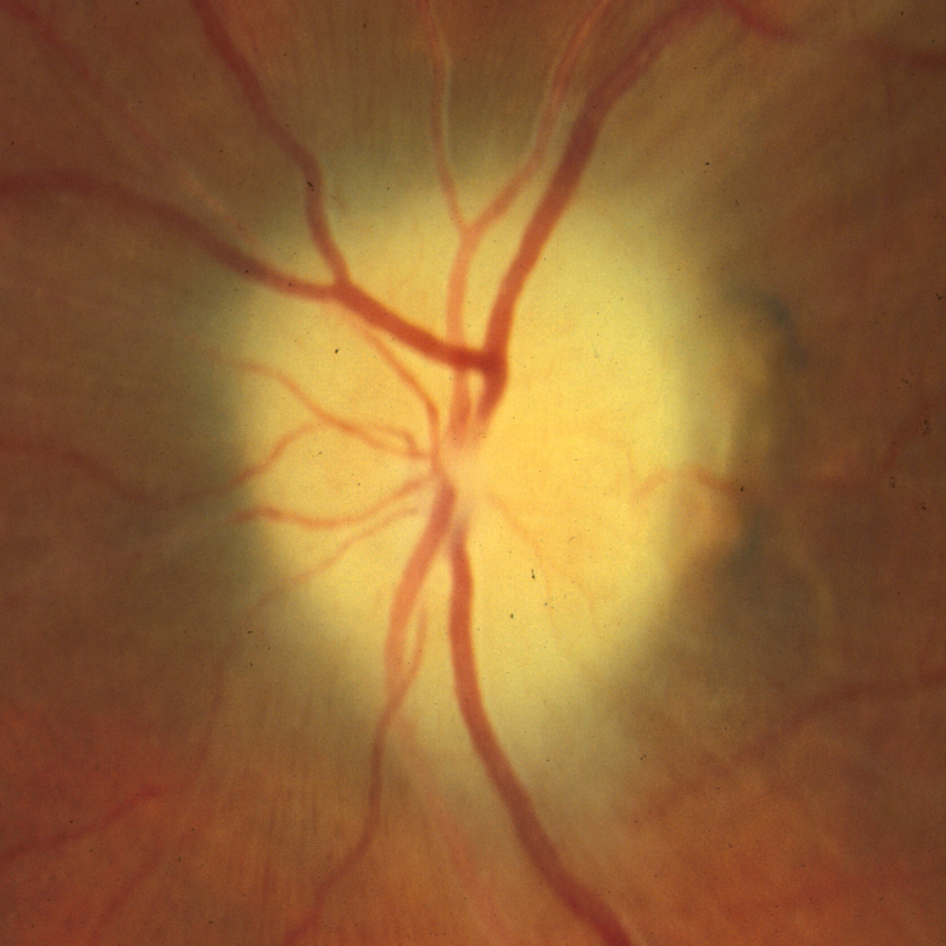
Primary (simple) atrophy follows damage behind the globe without previous disc swelling.
Typical causes include compression, retrobulbar optic neuritis, trauma and inherited neuropathies.
The disc looks chalky white with sharp, clean margins.
In addition, the lamina cribrosa often stays visible within the cup.
Fewer small vessels cross the disc margin, a finding known as the Kestenbaum sign.
Fundus Explorer Pro
Photograph the retinal findings described here with the phone already in your pocket — 22 D optics and built-in illumination in one handheld unit.
From Choroida — the team behind this siteSecondary Optic Atrophy
Secondary atrophy follows chronic disc swelling, such as long-standing papilledema or papillitis.
Glial tissue then fills the cup and blurs the disc margin.
As a result, the disc looks gray or dirty white, and the lamina is hidden.
Consecutive Optic Atrophy
Consecutive atrophy follows widespread retinal or choroidal disease that destroys ganglion cells.
Retinitis pigmentosa, central retinal artery occlusion and extensive panretinal photocoagulation are classic examples.
Here the disc often shows a waxy yellow pallor with attenuated arterioles.
Therefore, examine the retinal periphery before ordering neuroimaging for a pale disc.
Glaucomatous Atrophy
Glaucoma produces cupping with progressive loss of the neuroretinal rim.
The remaining rim usually keeps its pink color.
In contrast, pallor of the rim itself suggests a non-glaucomatous cause.
Ascending and Descending Optic Atrophy Types
Atrophy can also be grouped by the direction of degeneration.
Ascending atrophy starts in the retina or disc, as in glaucoma and consecutive atrophy.
Descending atrophy starts behind the globe and travels back toward the eye.
Because of this, a chiasmal lesion can eventually whiten the disc.
Pallor Patterns in Optic Atrophy Types
- Temporal pallor: papillomacular bundle loss, so think toxic, nutritional, hereditary or demyelinating causes
- Bow-tie (band) pallor: nasal and temporal sectors, which suggests chiasmal or optic tract damage
- Sectoral (altitudinal) pallor: superior or inferior sector, typical after ischemic optic neuropathy
- Diffuse pallor: whole disc, for example after compression or trauma
- Waxy pallor with thin vessels: retinal dystrophy or vascular occlusion
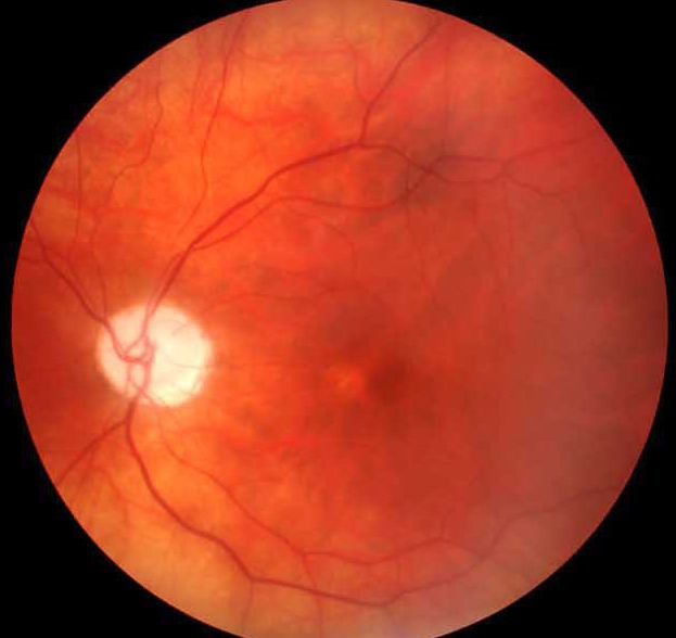
Temporal Pallor
The papillomacular bundle enters the temporal side of the disc.
Damage to this bundle causes central or cecocentral scotomas.
For example, toxic and nutritional neuropathy and dominant optic atrophy favor this pattern.
The temporal disc is normally slightly paler, so compare both eyes carefully.
Bow-Tie Atrophy
Bow-tie pallor affects the nasal and temporal disc while sparing the poles.
It develops in the eye that loses its temporal field, because nasal retinal fibers cross in the chiasm.
With an optic tract lesion, the contralateral eye shows band atrophy.
Meanwhile, the ipsilateral eye shows pallor at the superior and inferior poles.
Exam Technique for Optic Atrophy Types
Judge disc color with the same light source, and compare both eyes.
Pseudophakia and high myopia can make a disc look paler than it is.
Also check for a relative afferent pupillary defect in asymmetric disease.
Red-free light then reveals slit-like nerve fiber layer defects.
OCT and Field Testing
OCT measures retinal nerve fiber layer and ganglion cell layer thickness.
In some neuropathies, ganglion cell thinning may appear before obvious pallor.
In addition, field patterns help localize the lesion.
However, OCT alone cannot identify the cause.
When Optic Atrophy Types Need Imaging
Unexplained primary atrophy calls for orbital and cranial MRI with contrast, because compression must be excluded.
Progressive unilateral pallor and a pale rim with cupping also justify imaging.
Similarly, a field defect that respects the vertical meridian needs investigation.
For bilateral temporal pallor, add nutritional and toxic screening.
Counseling Patients With Optic Atrophy Types
Lost axons do not regenerate, so the pallor itself will not reverse.
However, treating the cause can preserve the remaining vision.
Explain that the term describes the disc appearance, while the cause needs separate workup.
Finally, refer for low vision support when daily function is limited.
Pitfalls to Avoid
- Calling a pale disc glaucoma without checking rim color
- Skipping the retinal periphery in a patient with waxy pallor
- Diagnosing pallor in a pseudophakic eye without comparing the fellow eye
- Assuming a normal disc excludes a recent optic nerve injury


Document what you see
Two smartphone imaging tools built for everyday clinic use — one for the slit lamp, one for the fundus.
From Choroida — the team behind this siteReferences
- Bowling B. Kanski’s Clinical Ophthalmology: A Systematic Approach. 8th ed. Elsevier; 2016.
- American Academy of Ophthalmology. Basic and Clinical Science Course, Section 5: Neuro-Ophthalmology. San Francisco: AAO; 2023.
- Trobe JD, Glaser JS, Cassady J, Herschler J, Anderson DR. Nonglaucomatous excavation of the optic disc. Arch Ophthalmol. 1980;98:1046-1050.
- Greenfield DS, Siatkowski RM, Glaser JS, Schatz NJ, Parrish RK. The cupped disc: who needs neuroimaging? Ophthalmology. 1998;105:1866-1874.

Writes and reviews the clinical articles on Educate. Every post is checked against peer-reviewed sources before it goes live.