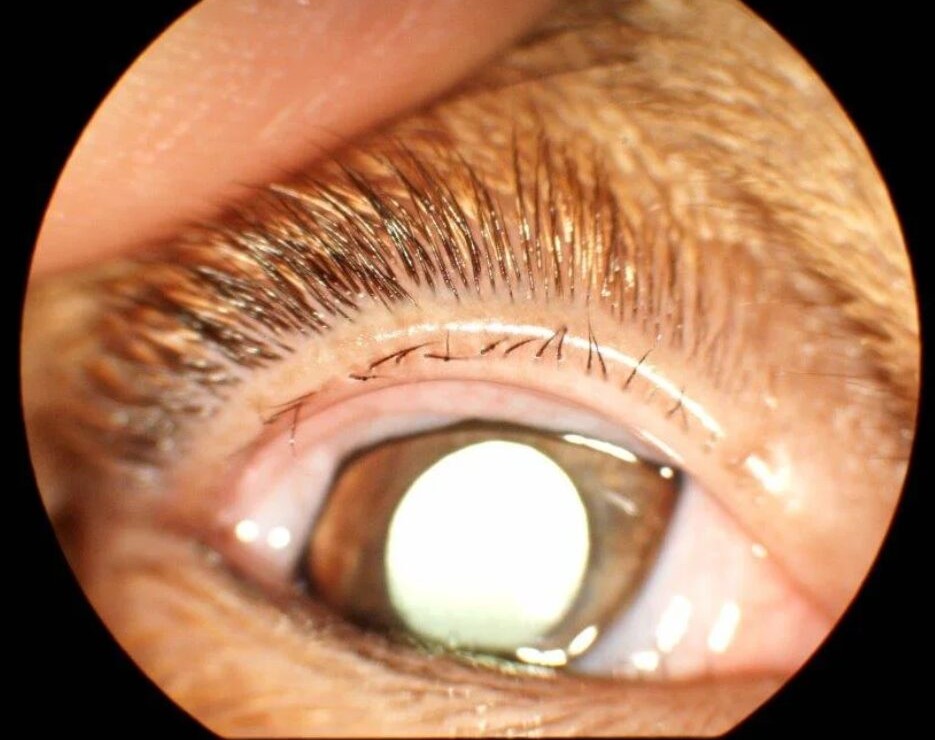
Ectopic cilia are lashes that grow through the tarsal conjunctiva instead of emerging from the lid margin.
They usually appear on the upper lid and point straight at the cornea, so every blink drags the lash across the epithelium. The condition is rare in humans, and most published descriptions are single case reports. However, it is easy to miss because the lid margin looks normal.
What Ectopic Cilia Are
An ectopic cilium arises from a follicle that sits abnormally deep in the tarsus, and the shaft then grows backward and exits through the palpebral conjunctiva. Most reports describe a single lash or a small tuft, and the exit point usually lies a few millimeters above the upper lid margin.
Origin of Ectopic Cilia
The exact mechanism is uncertain, but many authors consider it a congenital developmental error because cases often present in childhood. Lash follicles and meibomian glands share an embryologic origin, so a misplaced follicle can develop within the tarsal plate.
All-fit Slit-Lamp Adapter
Record and share exactly what you see at the slit lamp. One adapter fits any slit lamp or surgical microscope — and any smartphone.
From Choroida — the team behind this siteSymptoms Patients Report
Patients describe a sharp foreign body sensation that worsens with blinking, and tearing, photophobia and redness are also common. Because symptoms return after each “foreign body removal,” many patients see several doctors before diagnosis.
Corneal Damage From Ectopic Cilia
The lash scratches the superior cornea with each blink, and fluorescein therefore shows vertical linear abrasions, similar to a retained subtarsal foreign body. Repeated trauma can then cause punctate keratopathy, recurrent erosions or superficial scarring, and a chronic epithelial defect may become infected (see stromal corneal ulcers).
Examining for Ectopic Cilia
First, stain the cornea and note the pattern of damage, and next evert the upper lid and inspect the tarsal conjunctiva under high magnification. The lash may be short, fine and unpigmented, so it hides easily.
A small conjunctival papule or a dark dot at the exit site can be the only clue. Finally, gently sweep a cotton tip across the tarsus, since patients often feel pain at the exact spot.
Ectopic Cilia vs Distichiasis
Distichiasis is an extra row of lashes that emerge from the meibomian gland orifices, and these lashes therefore exit on the lid margin, behind the normal row. Ectopic cilia, by contrast, exit through the conjunctiva itself, away from the margin. Distichiasis also often involves several lashes, while ectopic cilia are usually single.
Ectopic Cilia vs Trichiasis
In trichiasis, the lashes arise from their normal position in the anterior lamella, but they grow toward the globe because of scarring or follicle damage (see trichiasis). Ectopic cilia, in contrast, have an abnormal exit site on the inner lid surface.
Other Conditions to Exclude
- Subtarsal foreign body
- Entropion with lash contact
- Epiblepharon in children
- Superior limbic keratoconjunctivitis
- Floppy eyelid syndrome
Why Ectopic Cilia Are Missed
The lid margin looks normal on routine examination, and examiners rarely evert the lid when there is no history of a foreign body. A fine lash can also break during eversion and leave only a stump, so unexplained superior linear staining should always prompt careful eversion.
Temporary Relief in Ectopic Cilia
Simple epilation with fine forceps gives immediate comfort, but the follicle remains, so the lash regrows within weeks. Lubricants and a bandage contact lens can protect the cornea while surgery is planned. Treat any epithelial defect with a topical antibiotic, as for other abrasions (see corneal abrasion).
Definitive Removal of Ectopic Cilia
Definitive treatment removes the follicle, so surgeons usually excise a small block of tarsal conjunctiva and tarsus around the lash under local anesthesia. The excision must include the entire follicle because a remnant can regrow.
Electrolysis or radiofrequency ablation has also been described for a single lash, but the deep follicle may be destroyed incompletely. Sending the tissue for histology confirms the diagnosis and the completeness of excision.
Risks of Surgery
Complications are uncommon after a small excision, and possible problems include bleeding, granuloma, tarsal notching and recurrence.
Counseling the Patient With Ectopic Cilia
Explain that the lash grows from a misplaced root inside the lid, and that pulling it out helps for a short time, but regrowth is expected. Surgery offers the best chance of cure, although recurrence remains possible. Also, warn parents that young children may need general anesthesia for the procedure.
Follow-Up After Removal
Review the patient after one to two weeks to check healing and corneal staining, then evert the lid again at later visits to detect any new lash. Persistent staining should prompt a search for a second lash or another cause.
Pitfalls to Avoid
- Do not label recurrent superior abrasions as erosions without everting the lid
- Never rely on epilation alone as definitive treatment
- Check the same lid for a second ectopic lash


Document what you see
Two smartphone imaging tools built for everyday clinic use — one for the slit lamp, one for the fundus.
From Choroida — the team behind this siteReferences
- Bowling B. Kanski’s Clinical Ophthalmology: A Systematic Approach. 8th ed. Elsevier; 2016.
- American Academy of Ophthalmology. Basic and Clinical Science Course, Section 7: Oculofacial Plastic and Orbital Surgery. San Francisco: AAO; 2023.
- American Academy of Ophthalmology. Basic and Clinical Science Course, Section 8: External Disease and Cornea. San Francisco: AAO; 2023.
- Ferreira IS, Bernardes TF, Bonfioli AA. Trichiasis. Semin Ophthalmol. 2010;25:66-71.
- Yanoff M, Duker JS. Ophthalmology. 5th ed. Elsevier; 2019.

Writes and reviews the clinical articles on Educate. Every post is checked against peer-reviewed sources before it goes live.