Lymphedema distichiasis syndrome is an autosomal dominant disorder that combines an extra row of eyelashes with primary limb lymphedema.
Most families carry a pathogenic variant in FOXC2, a forkhead transcription factor gene on chromosome 16q24.
Because the eye findings often appear first, the ophthalmologist may make the initial diagnosis.
What Lymphedema Distichiasis Is
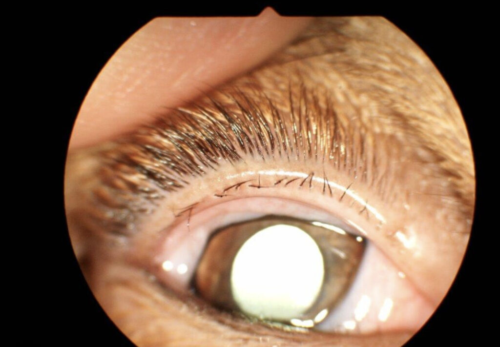
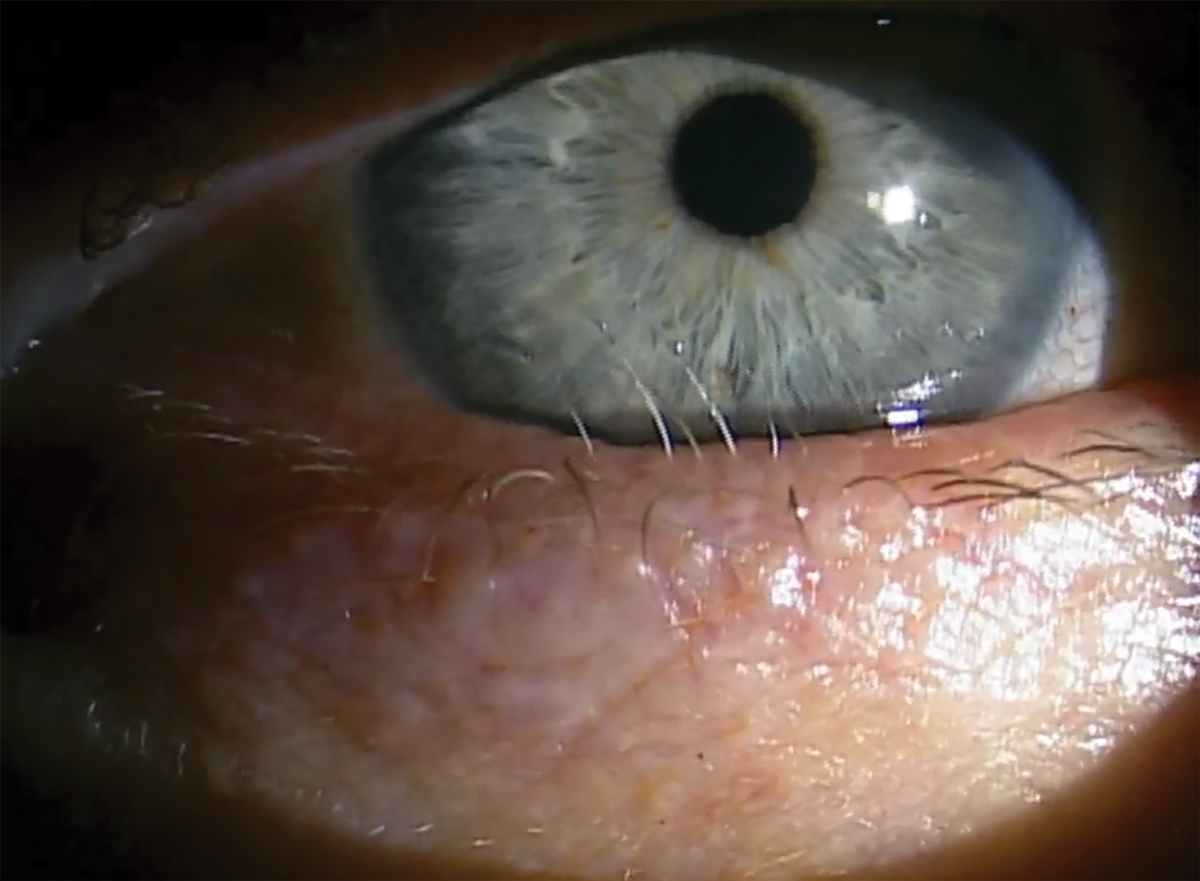
Distichiasis means a second row of lashes emerging from the posterior lid margin.
In this syndrome, the extra lashes grow from meibomian gland orifices or just behind them.
The leg swelling, by contrast, reflects abnormal lymphatic and venous valve development.
Therefore, one gene links two very different complaints.
Genetics of Lymphedema Distichiasis
Penetrance is high, although expression varies within one pedigree.
For example, a parent may have only lashes while a child develops marked swelling.
As a result, a careful family history often reveals affected relatives who never sought care.
All-fit Slit-Lamp Adapter
Record and share exactly what you see at the slit lamp. One adapter fits any slit lamp or surgical microscope — and any smartphone.
From Choroida — the team behind this siteWho Presents and When
Distichiasis is often present at birth or noticed in early childhood.
Children may rub their eyes, avoid light or show chronic redness.
Lymphedema usually starts later, often around puberty or in early adulthood.
Then it typically affects the lower limbs, sometimes asymmetrically.
Lid Findings at the Slit Lamp
Examine the lid margin with high magnification and good illumination.
The accessory lashes sit posterior to the gray line, in the meibomian orifice row.
They are often thinner, shorter and less pigmented than normal cilia.
In addition, some lashes point toward the globe while others lie flat along the margin.
Count them on all four lids, because involvement is usually bilateral but uneven.
Meibomian Glands in Lymphedema Distichiasis
The extra lashes develop where meibomian glands should form.
Consequently, gland expression yields little secretion in involved segments.
Meibography, when available, may show reduced or absent glands.
Therefore, evaporative dry eye can coexist with mechanical irritation.
Corneal Risk in Lymphedema Distichiasis
Lashes rubbing the cornea cause punctate epithelial erosions, usually inferiorly.
With time, patients may develop recurrent abrasions, pannus or scarring.
Any epithelial defect also raises the chance of infection (see microbial keratitis risk factors).
For this reason, stain every eye with fluorescein, even when symptoms seem mild.
Some patients adapt and report few complaints despite clear staining.
Systemic Associations of Lymphedema Distichiasis
Ask about the following, because they change counseling and referral:
- Lower limb lymphedema, often from puberty onward
- Early onset varicose veins from venous valve failure
- Ptosis
- Congenital heart disease
- Cleft palate
- Spinal extradural cysts
- Recurrent cellulitis in swollen limbs
Differential Diagnosis
Acquired distichiasis follows chronic cicatrizing disease, such as ocular cicatricial pemphigoid, Stevens-Johnson syndrome or chemical injury.
Trichiasis involves misdirected lashes from the normal anterior row.
Epiblepharon pushes normal lashes against the cornea in children.
Isolated congenital distichiasis without lymphedema also exists.
However, FOXC2 testing can reveal the syndrome in apparently isolated cases.
Confirming the Diagnosis
Diagnosis rests on lid examination plus family and limb history.
Genetic testing for FOXC2 confirms the syndrome and also helps relatives.
Then refer to a lymphedema service or genetics clinic when testing is positive.
Conservative Lash Management in Lymphedema Distichiasis
Start with lubrication, preferably preservative-free drops and a night ointment.
Mechanical epilation gives quick relief, because it removes the offending lash.
However, lashes regrow within weeks and often feel coarser.
Meanwhile, a bandage contact lens can protect the cornea while you plan definitive care.
Ablative Treatment
Electrolysis destroys individual follicles and suits a small number of lashes.
Radiofrequency and argon laser ablation work in a similar way.
Recurrence is common, so patients often need several sessions.
In addition, scarring of the margin can follow repeated treatment.
Surgical Options in Lymphedema Distichiasis
Extensive distichiasis may need lid splitting at the gray line.
The surgeon then treats the posterior lamella follicles with cryotherapy or direct excision.
Cryotherapy can cause depigmentation, notching and further meibomian loss.
Therefore, apply it to the posterior lamella only and protect the normal anterior row.
Counseling the Patient
Explain that no treatment guarantees permanent lash removal.
Also discuss the dominant inheritance and a 50% risk for each child.
Encourage relatives to have a lid examination, even without symptoms.
Finally, patients with leg swelling should report cellulitis early.
Follow-Up in Lymphedema Distichiasis
Review untreated patients for staining and new lashes.
After ablation, check for regrowth, lid notching and worsening dry eye.
Children need closer review, because they rarely report corneal symptoms reliably.
Pitfalls to Avoid
- Do not dismiss fine pale lashes, because they still abrade the cornea
- Never treat the anterior lash row with cryotherapy
- Do not miss the lymphedema history in a child labeled with trichiasis
- Remember that dry eye persists after successful lash removal


Document what you see
Two smartphone imaging tools built for everyday clinic use — one for the slit lamp, one for the fundus.
From Choroida — the team behind this siteReferences
- Fang J, Dagenais SL, Erickson RP, et al. Mutations in FOXC2 (MFH-1), a forkhead family transcription factor, are responsible for the hereditary lymphedema-distichiasis syndrome. Am J Hum Genet. 2000;67:1382-1388.
- Brice G, Mansour S, Bell R, et al. Analysis of the phenotypic abnormalities in lymphoedema-distichiasis syndrome in 74 patients with FOXC2 mutations or linkage to 16q24. J Med Genet. 2002;39:478-483.
- Mellor RH, Brice G, Stanton AW, et al. Mutations in FOXC2 are strongly associated with primary valve failure in veins of the lower limb. Circulation. 2007;115:1912-1920.
- Bowling B. Kanski’s Clinical Ophthalmology: A Systematic Approach. 8th ed. Elsevier; 2016.

Writes and reviews the clinical articles on Educate. Every post is checked against peer-reviewed sources before it goes live.