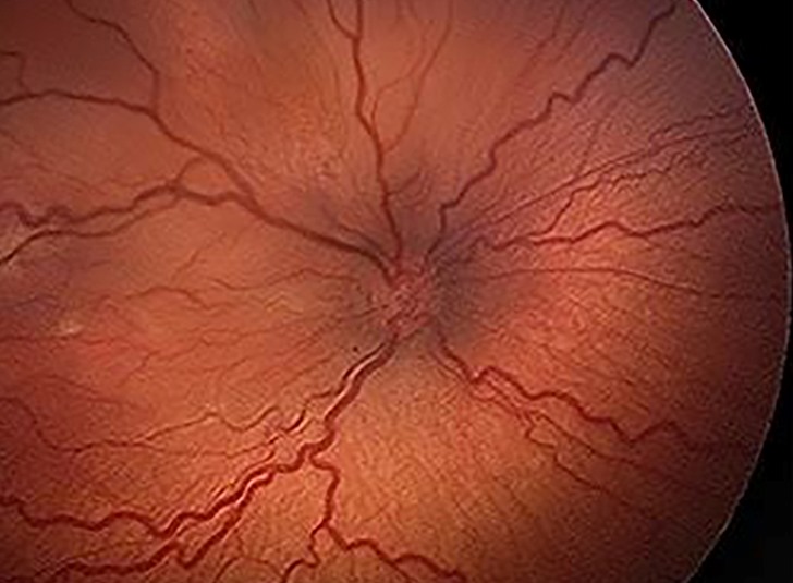
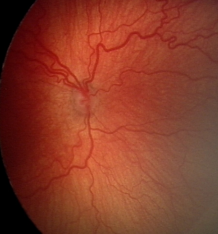
Plus disease is abnormal dilation and tortuosity of the posterior retinal vessels in retinopathy of prematurity (ROP).
It reflects disease activity, separate from zone and stage.
Therefore, it is often the sign that decides whether an infant needs treatment this week.
What Plus Disease Means
ICROP3 defines plus disease by the appearance of vessels within zone I.
Arteries become tortuous, while veins become dilated.
The change reflects increased flow and shunting through abnormal vessels at the junction of vascular and avascular retina.
As a result, the posterior pole carries a readable signal of peripheral activity.
Plus Disease as a Spectrum
Older classifications treated plus as simply present or absent.
However, ICROP3 describes a continuum from normal vessels through pre-plus to plus.
Vascular changes can also progress or regress between visits.
Therefore, record the direction of change at every examination.
Fundus Explorer Pro
Photograph the retinal findings described here with the phone already in your pocket — 22 D optics and built-in illumination in one handheld unit.
From Choroida — the team behind this sitePre-Plus vs Plus Disease
Pre-plus shows more dilation and tortuosity than normal, yet not enough for plus.
The 2005 ICROP revision introduced this category.
Pre-plus alone does not meet treatment criteria.
However, it signals higher risk and usually shortens the follow-up interval.
From Quadrants to Zone I
The CRYO-ROP trial originally required vessel changes in at least two quadrants.
Later, ICROP3 moved away from counting quadrants.
Examiners now judge the overall appearance of vessels within zone I.
Because peripheral vessels vary widely, the central view gives a more consistent reference.
How Examiners Judge Plus Disease
Most examiners use binocular indirect ophthalmoscopy with a 28 or 20 diopter lens.
First, they assess the arteries for tortuosity.
Next, they assess the veins for dilation.
Then they compare the overall impression with the standard reference photographs.
Arterial Tortuosity in Plus Disease
Studies of expert grading suggest that arterial tortuosity strongly influences the final call.
However, mild tortuosity can occur in healthy preterm infants (see retinal vascular tortuosity).
Therefore, judge it against the reference standard, not against an adult fundus.
Venous Dilation
Veins widen as flow through the shunts increases.
Dilation is easier to underestimate when the view is small or hazy.
In addition, a single dilated vein carries less weight than a generalized change.
Why Plus Disease Drives Treatment
The ETROP trial showed benefit from treating high-risk eyes earlier than the older threshold.
As a result, plus disease became central to the definition of type 1 ROP:
- Zone I, any stage, with plus disease
- Also zone I, stage 3, without plus disease
- Finally, zone II, stage 2 or 3, with plus disease
Type 2 ROP and Observation
Type 2 ROP includes zone I stage 1 or 2 without plus, and zone II stage 3 without plus.
These eyes usually receive close observation.
However, the appearance of plus converts many of them to type 1.
Timing of Treatment
Screening guidance advises treating type 1 ROP generally within 72 hours of diagnosis.
Therefore, a new finding of plus should trigger same-day communication with the treating team.
Delay allows fibrovascular proliferation to advance toward detachment (see ROP diagnosis and management).
Plus Disease in Aggressive ROP
ICROP3 renamed aggressive posterior ROP as aggressive ROP (A-ROP).
In A-ROP, plus disease is often severe and appears out of proportion to peripheral findings.
Also, the classic stage sequence may not occur (see aggressive posterior ROP).
Regression After Treatment
After laser or anti-VEGF injection, plus disease usually improves before peripheral changes settle.
However, reactivation can occur later, and returning dilation may be an early clue.
Therefore, extended follow-up is needed after injection (see laser versus anti-VEGF).
Interobserver Variability
Experts do not always agree on plus disease.
For example, Chiang and colleagues found imperfect agreement even among experienced graders.
As a result, borderline eyes benefit from a second opinion or serial imaging.
Imaging and Automated Grading of Plus Disease
Wide-field imaging allows side-by-side comparison across visits.
In addition, deep learning systems have shown high agreement with expert consensus in research settings.
Still, these tools support clinical judgment and do not replace the bedside examination.
Counseling Parents
Explain that the retinal vessels show how active the disease is.
Then describe why treatment may be needed before any visible scarring.
Also, stress that follow-up continues after treatment, because reactivation remains possible.
Pitfalls to Avoid
- Calling plus from peripheral vessels alone
- Ignoring pre-plus when setting the next visit
- Missing plus through a small pupil or hazy media
- Assuming quick improvement after anti-VEGF means cure


Document what you see
Two smartphone imaging tools built for everyday clinic use — one for the slit lamp, one for the fundus.
From Choroida — the team behind this siteReferences
- Chiang MF, Quinn GE, Fielder AR, et al. International Classification of Retinopathy of Prematurity, Third Edition. Ophthalmology. 2021;128:e51-e68.
- International Committee for the Classification of Retinopathy of Prematurity. The International Classification of Retinopathy of Prematurity revisited. Arch Ophthalmol. 2005;123:991-999.
- Early Treatment for Retinopathy of Prematurity Cooperative Group. Revised indications for the treatment of retinopathy of prematurity: results of the ETROP randomized trial. Arch Ophthalmol. 2003;121:1684-1694.
- Chiang MF, Jiang L, Gelman R, et al. Interexpert agreement of plus disease diagnosis in retinopathy of prematurity. Arch Ophthalmol. 2007;125:875-880.
- Fierson WM, American Academy of Pediatrics Section on Ophthalmology, et al. Screening examination of premature infants for retinopathy of prematurity. Pediatrics. 2018;142:e20183061.
- Brown JM, Campbell JP, Beers A, et al. Automated diagnosis of plus disease in retinopathy of prematurity using deep convolutional neural networks. JAMA Ophthalmol. 2018;136:803-810.

Writes and reviews the clinical articles on Educate. Every post is checked against peer-reviewed sources before it goes live.