Chorioretinal scars are focal areas where inflammation, injury or degeneration has destroyed retina, pigment epithelium and choroid together.
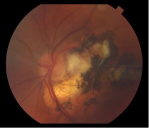
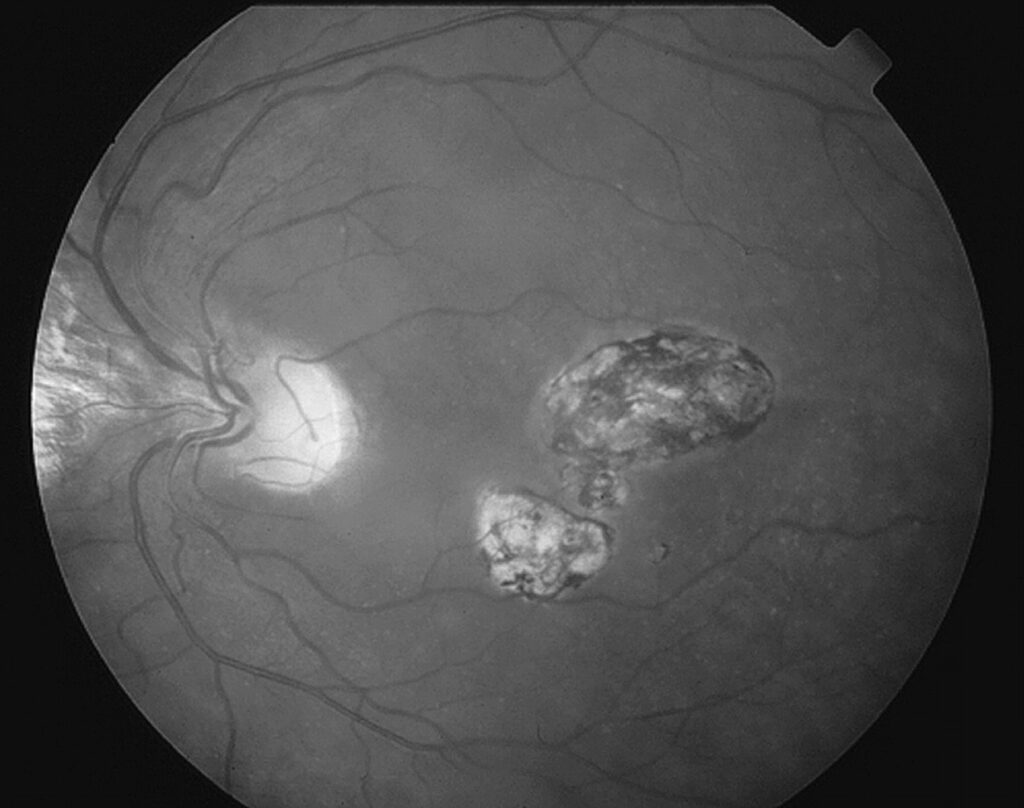
On fundus examination, they appear as white atrophic patches, pigmented plaques, or both.
Because several causes can reactivate, the pattern guides both diagnosis and follow-up.
Therefore, read the shape, number, location and pigment before ordering tests.
What Chorioretinal Scars Look Like
First, loss of pigment epithelium and choriocapillaris exposes white sclera or large choroidal vessels.
At the same time, surviving pigment cells migrate and clump at the scar margin.
As a result, most scars show a pale center with a dark rim.
Toxoplasmosis Chorioretinal Scars
Toxoplasma scars are usually single or few, round to oval, and deeply pigmented.
Also, they often sit in the posterior pole, and congenital lesions commonly involve the macula.
Clustered satellite scars may mark previous recurrences.
However, once the disease is quiet, the vitreous is usually clear.
Fundus Explorer Pro
Photograph the retinal findings described here with the phone already in your pocket — 22 D optics and built-in illumination in one handheld unit.
From Choroida — the team behind this siteReactivation in Toxoplasma Scars
Recurrence typically appears as a fluffy white focus at the border of an old scar.
Then dense vitritis blurs the view, producing the classic “headlight in the fog” appearance.
In addition, patients report floaters and blurred vision, sometimes with redness and photophobia.
Also, a full checklist is in toxoplasmosis scar reactivation.
Histo-Like Punched-Out Spots
Presumed ocular histoplasmosis syndrome produces small, round, atrophic “histo spots” in the midperiphery and posterior pole.
In addition, peripapillary atrophy and linear peripheral streaks may appear.
However, the vitreous stays clear, which helps separate it from active uveitis.
Most patients have no symptoms until the macula becomes involved.
Multifocal Choroiditis and PIC
Multifocal choroiditis and punctate inner choroidopathy leave similar punched-out scars.
However, these patients are often young, myopic women.
Also, multifocal choroiditis may show anterior chamber or vitreous cells, while PIC lesions are smaller and cluster posteriorly.
Therefore, history and inflammation help separate them from histoplasmosis.
Reactivation in Punched-Out Chorioretinal Scars
First, the main threat is choroidal neovascularization arising at the edge of a macular scar.
Then look for a gray-green membrane, subretinal hemorrhage or a pigment ring.
Patients notice distortion, a new central spot, or sudden blur.
Also, new active inflammatory lesions look creamy yellow with soft borders.
Laser Chorioretinal Scars
Laser scars follow a geometry that disease rarely produces.
For example, panretinal photocoagulation leaves rows of uniform, pigmented spots that spare the posterior pole.
In addition, barrier laser forms rings around a break or lattice (see laser retinopexy).
However, macular laser scars can enlarge over time, a process called atrophic creep.
Traumatic Choroidal Rupture
Blunt trauma can tear Bruch membrane, choriocapillaris and pigment epithelium.
The resulting scar is a curved white crescent concentric to the optic disc.
However, early subretinal hemorrhage may hide the rupture.
Then, as the blood clears, the yellow-white crescent becomes visible.
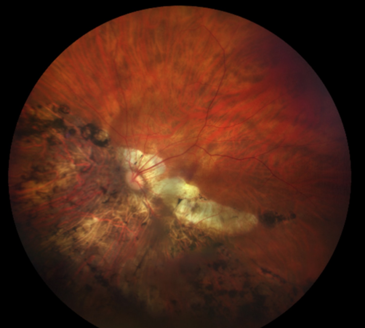
Trauma Chorioretinal Scars at Follow-Up
Choroidal neovascularization may grow from a rupture months or years later.
Therefore, advise trauma patients to report new distortion promptly.
Ruptures through the fovea also limit vision even without complications.
Myopic Chorioretinal Scars
High myopia produces patchy atrophy, lacquer cracks and peripapillary atrophy.
For example, patchy atrophy appears as well-defined gray-white areas, often near the disc or within a staphyloma.
In addition, a pigmented macular lesion may mark old neovascularization (see Fuchs spot).
Also, large atrophic areas often expand slowly over years.
Reactivation in Myopic Eyes
New myopic neovascularization usually presents as a small gray lesion with little fluid.
However, hemorrhage may also come from a fresh lacquer crack without any membrane.
Therefore, OCT and sometimes angiography help tell them apart.
As a result, confirmed membranes generally warrant prompt anti-VEGF treatment.
Other Causes to Consider
- Syphilis and tuberculosis, which can also leave multifocal scars
- Acute retinal necrosis, with scalloped peripheral atrophy
- Toxocara, often a single granuloma with a retinal fold
- Healed CMV retinitis in immunosuppressed patients
Imaging Chorioretinal Scars
First, fundus autofluorescence shows scars as dark areas with sharp borders.
In contrast, a bright rim may signal activity or ongoing pigment epithelium stress.
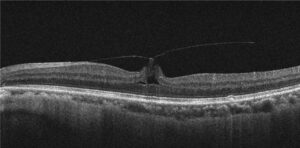
Next, OCT shows retinal thinning, RPE loss and increased signal transmission into the choroid.
Fluorescein angiography reveals neovascularization through early hyperfluorescence with late leakage, while inactive scars only stain.
Counseling Patients With Chorioretinal Scars
Most scars are stable and need no treatment.
However, patients should check each eye separately with an Amsler grid or near text.
In addition, explain that new distortion, floaters or a dark spot needs review within days.
Finally, treatment targets complications, because scar tissue cannot regenerate retina.
Pitfalls to Avoid
- Do not label every pigmented scar toxoplasmosis without checking the pattern
- Never miss fluid or blood at a scar edge, because it may signal neovascularization
- Do not treat quiet old scars with steroids or antimicrobials, because they are inactive
- Also, do not skip OCT when vision drops in a scarred eye


Document what you see
Two smartphone imaging tools built for everyday clinic use — one for the slit lamp, one for the fundus.
From Choroida — the team behind this siteReferences
- Holland GN. Ocular toxoplasmosis: a global reassessment. Part II: disease manifestations and management. Am J Ophthalmol. 2004;137:1-17.
- Macular Photocoagulation Study Group. Argon laser photocoagulation for neovascular maculopathy: three-year results from randomized clinical trials. Arch Ophthalmol. 1986;104:694-701.
- Ohno-Matsui K, Kawasaki R, Jonas JB, et al. International photographic classification and grading system for myopic maculopathy. Am J Ophthalmol. 2015;159:877-883.
- American Academy of Ophthalmology. Basic and Clinical Science Course, Section 12: Retina and Vitreous. San Francisco: AAO; 2023.
- Bowling B. Kanski’s Clinical Ophthalmology: A Systematic Approach. 8th ed. Elsevier; 2016.

Writes and reviews the clinical articles on Educate. Every post is checked against peer-reviewed sources before it goes live.