Superficial punctate keratitis (SPK) is a clinical sign of fine epithelial damage, and it has no single cause.
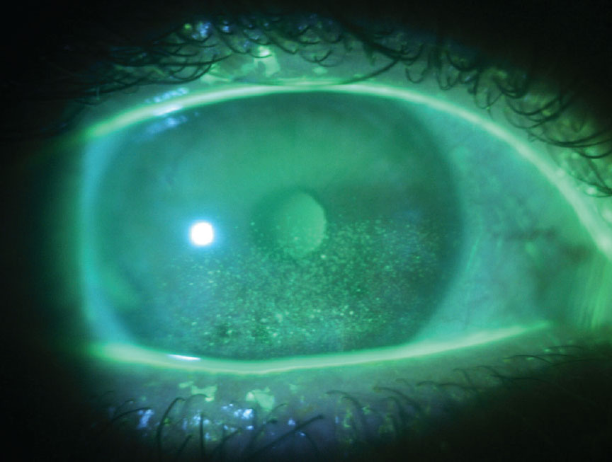
The slit lamp shows pinpoint lesions that stain with fluorescein across part or all of the cornea.
Therefore, the location of the staining often narrows the differential before any test is ordered.
What Superficial Punctate Keratitis Means
The term describes a pattern of epithelial injury, so it needs a cause attached.
Punctate epithelial erosions appear as tiny depressed defects that stain brightly with fluorescein.
In contrast, punctate epithelial keratitis shows small, swollen, gray-white cells that are visible before staining.
Symptoms of Superficial Punctate Keratitis
Patients usually report foreign body sensation, burning, tearing, and mild photophobia.
Vision may blur when lesions cross the visual axis.
However, symptoms often track poorly with staining, particularly in dry eye.
All-fit Slit-Lamp Adapter
Record and share exactly what you see at the slit lamp. One adapter fits any slit lamp or surgical microscope — and any smartphone.
From Choroida — the team behind this siteHow to Examine the Stain
First, check corneal sensation before you instill any anesthetic drop.
Next, apply a small amount of fluorescein and view the cornea with cobalt blue light.
Then lift the upper lid, because superior disease hides easily.
Our guide to corneal staining patterns reviews this map in more detail.
Inferior Superficial Punctate Keratitis
Inferior staining points first to the lid margin and to incomplete closure.
Staphylococcal blepharitis often produces this pattern, so examine the lashes and meibomian glands closely.
Lagophthalmos also exposes the inferior cornea during sleep, and morning symptoms support this cause.
Interpalpebral Staining
A horizontal band within the open lid fissure suggests evaporative stress or direct exposure.
Dry eye is a common explanation, particularly when the adjacent conjunctiva also stains.
Lissamine green helps here because it highlights conjunctival damage.
Superior Superficial Punctate Keratitis
Superior staining directs attention to the upper tarsus and limbus.
For example, superior limbic keratoconjunctivitis causes bulbar injection and tarsal papillae, and thyroid eye disease often accompanies it.
Floppy eyelid syndrome also belongs here, so test how easily the upper lid everts.
Inferonasal Staining and Drop Toxicity
Medication toxicity often favors the inferior or inferonasal cornea, where instilled drops collect and drain.
Glaucoma drops, aminoglycosides, topical antivirals, and anesthetics are frequent culprits.
As a result, take a complete drop history, including over-the-counter products.
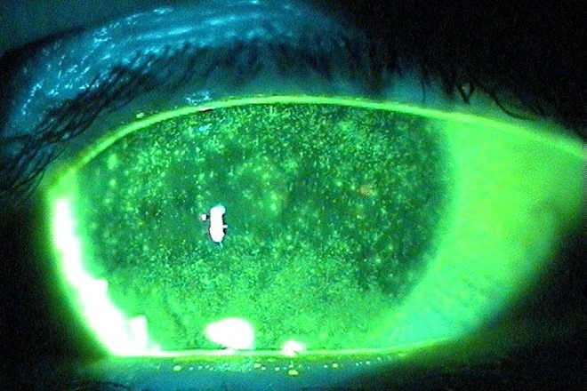
Diffuse Superficial Punctate Keratitis
Staining across the whole cornea suggests a widespread insult.
Adenoviral conjunctivitis is a typical cause, and follicles, a tender preauricular node, or a sick contact support it.
Contact lens solution reactions, chemical exposure, and severe aqueous deficiency can also stain diffusely.
Central Staining and Reduced Sensation
Central or inferocentral staining in a comfortable eye should raise concern for neurotrophic keratopathy.
Ask about prior herpes infection, diabetes, corneal surgery, and trigeminal injury.
Past herpes simplex keratitis is especially relevant, because repeated episodes can reduce corneal sensation.
Contact Lenses and Superficial Punctate Keratitis
Rigid lenses may cause peripheral staining at the 3 and 9 o’clock positions.
Soft lens solution reactions usually stain more diffusely, while overwear can affect the central cornea.
However, any infiltrate, significant pain, or anterior chamber reaction means infection must be excluded.
Mimics of Superficial Punctate Keratitis
Coarse, stellate, slightly elevated lesions in a quiet eye suggest Thygeson disease, a separate diagnosis.
Similarly, branching lesions with terminal bulbs point toward herpetic epithelial disease.
Early Acanthamoeba infection can also show irregular epithelial changes in a contact lens wearer.
Treating Superficial Punctate Keratitis
Treatment should address the cause, because lubricants alone relieve symptoms without correcting exposure, toxicity, or lid disease.
For example, lid hygiene helps blepharitis, while drop reduction helps toxicity.
Lens wearers should stop wear until the surface recovers.
Topical steroids can reduce inflammation, but they carry risks of worsening herpetic disease and raising intraocular pressure.
Preservative-Free Options in Superficial Punctate Keratitis
Preservative-free lubricants suit patients who need frequent dosing, because repeated exposure to preservatives can injure the epithelium.
In addition, a lubricating ointment at bedtime protects the cornea in lagophthalmos.
However, reassess after two to four weeks, since persistent staining despite these measures calls for a new look at the cause.
When to Order Further Testing
Most cases need no laboratory work, because the pattern and history usually settle the diagnosis.
However, consider viral PCR or culture when the epithelial lesions look dendritic or the course is atypical.
Also refer patients with suspected Sjögren disease or thyroid disease for systemic evaluation.
Follow-Up and Counseling
Compare the same corneal zones at each visit, using a grading scheme such as the Oxford scheme when possible.
If staining persists, revisit the pattern, drop list, and corneal sensation.
Meanwhile, advise patients to return promptly for worsening pain, reduced vision, or a white corneal spot.
Pitfalls to Avoid
- Do not label every case as dry eye without mapping the stain
- Never skip upper lid eversion in superior staining
- Do not prescribe chronic topical anesthetics for symptom relief
- Avoid empiric steroids before you exclude herpetic disease


Document what you see
Two smartphone imaging tools built for everyday clinic use — one for the slit lamp, one for the fundus.
From Choroida — the team behind this siteReferences
- Thygeson P. Superficial punctate keratitis. JAMA. 1950;144:1544-1549.
- Bron AJ, Evans VE, Smith JA. Grading of corneal and conjunctival staining in the context of other dry eye tests. Cornea. 2003;22:640-650.
- Wolffsohn JS, Arita R, Chalmers R, et al. TFOS DEWS II diagnostic methodology report. Ocul Surf. 2017;15:539-574.
- Baudouin C, Labbe A, Liang H, Pauly A, Brignole-Baudouin F. Preservatives in eyedrops: the good, the bad and the ugly. Prog Retin Eye Res. 2010;29:312-334.
- Bowling B. Kanski’s Clinical Ophthalmology: A Systematic Approach. 8th ed. Elsevier; 2016.

Writes and reviews the clinical articles on Educate. Every post is checked against peer-reviewed sources before it goes live.