Keratitis vs conjunctivitis is a common triage question because both conditions can produce a red, irritated eye.
Conjunctivitis usually follows a benign course, while keratitis can scar the cornea and threaten vision.
Therefore, the first examination should confirm or exclude corneal involvement.
Keratitis vs Conjunctivitis: Core Difference
Keratitis means inflammation of the cornea, with or without infection.
Conjunctivitis affects the membrane covering the sclera and lining the eyelids.
Because the cornea refracts light and has dense sensory innervation, corneal disease often causes pain and blur.
In contrast, conjunctivitis usually causes milder discomfort and spares vision.
Symptoms in Keratitis vs Conjunctivitis
Patients with keratitis often report pain, light sensitivity and difficulty opening the eye.
They may also describe blur that persists after blinking.
In contrast, conjunctivitis more often causes itching, burning, grittiness or morning lid crusting.
However, reduced corneal sensation can mask pain in neurotrophic keratitis.
All-fit Slit-Lamp Adapter
Record and share exactly what you see at the slit lamp. One adapter fits any slit lamp or surgical microscope — and any smartphone.
From Choroida — the team behind this siteDischarge and Laterality
Watery discharge after an upper respiratory infection suggests adenoviral conjunctivitis.
Itching with stringy mucus and bilateral symptoms favors allergic disease.
Purulent discharge suggests bacterial infection, and rapid, copious pus raises concern for gonococcal conjunctivitis.
However, bacterial keratitis can also produce discharge, so this sign cannot exclude corneal disease.
History That Raises Suspicion
Contact lens wear is a major risk factor for microbial keratitis, so ask about overnight use.
Water exposure also raises suspicion for Acanthamoeba keratitis, especially when pain seems severe for the findings.
Next, ask about trauma, plant matter, prior herpes, ocular surgery and topical steroid use.
Keratitis vs Conjunctivitis at the Slit Lamp
Begin with visual acuity, because a new decline points toward corneal or intraocular disease.
Then inspect the lids, conjunctiva, cornea and anterior chamber in a fixed order.
Diffuse injection that increases toward the fornices suggests conjunctivitis.
By contrast, circumlimbal ciliary flush suggests corneal or intraocular inflammation.
Corneal Signs in Keratitis
A stromal infiltrate appears as a white or gray opacity because leukocytes accumulate in the tissue.
Examiners may also see an epithelial defect, stromal edema, thinning or anterior chamber cells.
A hypopyon signals significant inflammation and calls for urgent management.
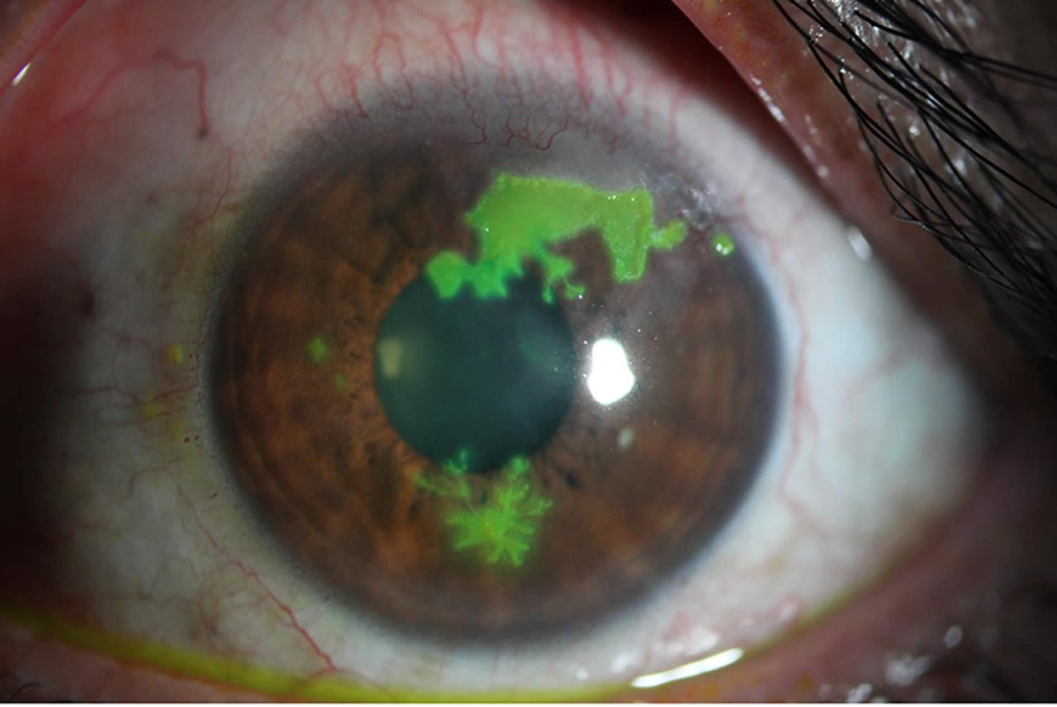
Fluorescein Staining in Keratitis vs Conjunctivitis
Instill fluorescein in any red eye with pain, blur or contact lens wear.
Then view the cornea under cobalt blue light.
A dendrite with terminal bulbs suggests herpes simplex epithelial keratitis.
In addition, a defect over a white infiltrate raises suspicion for microbial keratitis.
Punctate staining is less specific, because dry eye, drop toxicity and viral disease can all cause it.
When the Conditions Overlap
Adenovirus often involves both tissues, producing epidemic keratoconjunctivitis.
Early punctate keratitis may later give way to subepithelial infiltrates and persistent glare.
Similarly, gonococcal conjunctivitis can progress to corneal ulceration and perforation.
Therefore, a confirmed conjunctivitis still requires corneal review when symptoms change.
Triage: Keratitis vs Conjunctivitis
A structured red eye triage helps separate routine cases from urgent ones.
Arrange same-day ophthalmic assessment for significant pain, photophobia or reduced vision.
Contact lens wear with a red, painful eye also warrants same-day review.
The same applies to corneal opacity, hypopyon, recent trauma or copious purulent discharge.
Treating Conjunctivitis
Viral conjunctivitis usually needs supportive care, including cold compresses, lubricants and hand hygiene.
Antibiotics do not treat the virus, and they can add cost and surface toxicity.
Many bacterial cases resolve spontaneously, although topical antibiotics can speed recovery modestly.
Allergic disease often responds to topical antihistamine and mast cell stabilizer drops.
Treating Keratitis
Suspected bacterial keratitis needs frequent topical antibiotics, using a fluoroquinolone or fortified agents.
Herpetic, fungal and Acanthamoeba disease each require directed therapy, so the suspected cause guides treatment.
Treatment can sterilize the ulcer; however, it cannot always prevent scarring or visual loss.
Therefore, warn patients early about prolonged treatment and frequent visits.
Keratitis vs Conjunctivitis: Steroid Risk
Topical steroids can relieve redness and hide the warning signs of infection.
They can also worsen herpes simplex epithelial keratitis and fungal keratitis, while raising intraocular pressure.
In the SCUT trial, adjunctive steroids showed no overall visual benefit at three months in treated bacterial ulcers.
Thus, avoid steroids for a red eye until a slit lamp examination defines the diagnosis.
Counseling in Keratitis vs Conjunctivitis
Tell patients with infectious conjunctivitis to avoid sharing towels and to wash hands often.
Contact lens wearers should stop lens use and bring their lenses and case to the visit.
Finally, advise urgent return for pain, light sensitivity or blurred vision.
Pitfalls to Avoid
- Do not label a painful red eye as conjunctivitis without staining the cornea
- Do not overlook contact lens wear in a patient with discharge
- Never prescribe topical anesthetic drops for home use
- Avoid patching a suspected infected corneal lesion


Document what you see
Two smartphone imaging tools built for everyday clinic use — one for the slit lamp, one for the fundus.
From Choroida — the team behind this siteReferences
- Azari AA, Barney NP. Conjunctivitis: a systematic review of diagnosis and treatment. JAMA. 2013;310:1721-1729.
- Varu DM, Rhee MK, Akpek EK, et al. Conjunctivitis Preferred Practice Pattern. Ophthalmology. 2019;126:P94-P169.
- Lin A, Rhee MK, Akpek EK, et al. Bacterial Keratitis Preferred Practice Pattern. Ophthalmology. 2019;126:P1-P55.
- Austin A, Lietman T, Rose-Nussbaumer J. Update on the management of infectious keratitis. Ophthalmology. 2017;124:1678-1689.
- Srinivasan M, Mascarenhas J, Rajaraman R, et al. Corticosteroids for bacterial keratitis: the Steroids for Corneal Ulcers Trial (SCUT). Arch Ophthalmol. 2012;130:143-150.

Writes and reviews the clinical articles on Educate. Every post is checked against peer-reviewed sources before it goes live.