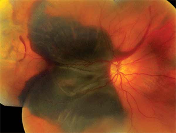
Sub-RPE hemorrhage is bleeding between the retinal pigment epithelium and Bruch’s membrane.
Because pigment covers the blood, the lesion can look gray, green or almost black.
However, careful imaging usually separates it from choroidal melanoma.
Why Sub-RPE Hemorrhage Looks Dark
The RPE masks the red color of fresh blood, so the examiner sees a dark elevated lesion.
In contrast, subretinal blood usually looks red because only the neurosensory retina covers it.
A rim of red subretinal blood around a dark center is therefore a useful clue.
Common Causes of Sub-RPE Hemorrhage
Neovascular AMD is a common source, especially type 1 macular neovascularization.
Polypoidal choroidal vasculopathy also produces large hemorrhagic pigment epithelial detachments.
In addition, a retinal arterial macroaneurysm can bleed at several levels, including beneath the RPE.
Peripheral lesions should raise suspicion of PEHCR.
Fundus Explorer Pro
Photograph the retinal findings described here with the phone already in your pocket — 22 D optics and built-in illumination in one handheld unit.
From Choroida — the team behind this siteWhat the Patient Reports
Patients with a bleed often describe sudden central blur, distortion or a dark spot.
Melanoma more often causes gradual symptoms, or none at all.
However, these histories overlap, so symptoms only set the level of suspicion.
Also ask about prior AMD, antithrombotic drugs and previous cancer.
Fundus Signs of Sub-RPE Hemorrhage
Examine both eyes first.
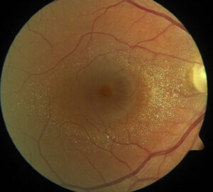
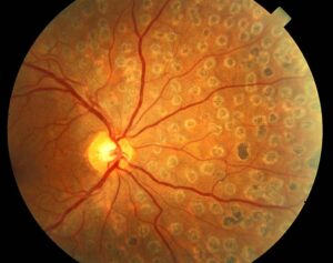
Drusen or a disciform scar in the fellow eye favors a bleed from AMD.
Exudates and adjacent red blood also support a vascular source.
Next, check the lesion margin, because polyps and neovascular tissue often lie at the edge.
Melanoma Features That Raise Concern
Orange pigment and overlying subretinal fluid increase concern for choroidal melanoma.
A break through Bruch’s membrane can also create a mushroom shape.
However, melanoma can bleed, so visible blood does not exclude a tumor.
OCT in Sub-RPE Hemorrhage
OCT shows a dome-shaped RPE elevation with reflective material beneath it.
Fresh blood often casts a dense shadow, which may hide the deeper choroid.
Therefore, search the margins for a double-layer sign, a notch or a polypoidal lesion.
Enhanced depth imaging can help in shallow lesions, because melanoma arises within the choroid itself.
Ultrasound in Sub-RPE Hemorrhage
Ultrasound often gives the most useful information when a lesion is thick.
Melanoma typically shows low to medium internal reflectivity on A-scan and acoustic hollowness on B-scan.
Choroidal excavation and vascular pulsation also support a tumor.
In contrast, a clot tends to look irregular and lacks true vascular pulsation.
However, organized blood can appear solid, so repeat the scan when findings conflict.
Angiography in Sub-RPE Hemorrhage
On fluorescein angiography, blood blocks fluorescence across the lesion.
Melanoma, by comparison, may show pinpoint leakage or a double circulation.
Indocyanine green reaches deeper, so it can reveal polyps or a neovascular network at the edge.
OCT angiography may also detect flow beside the bleed, although dense blood can block the signal.
Autofluorescence Clues
Fresh blood usually looks dark on autofluorescence.
As it breaks down, however, the signal may become bright.
Orange pigment in melanoma also glows because it contains lipofuscin.
Therefore, a bright signal alone proves little, and its evolution over time carries more weight.
How Sub-RPE Hemorrhage Changes Over Time
Serial imaging often settles the question.
Blood usually flattens and fades, often toward yellow, over weeks to months.
Melanoma, however, persists or grows.
For this reason, repeat photographs, OCT and ultrasound at a short interval when the diagnosis stays uncertain.
Other Lesions in the Differential
Shields and colleagues list hemorrhagic RPE detachment among the common pseudomelanomas.
Other mimics include choroidal nevus, congenital RPE hypertrophy and melanocytoma.
Most of these lesions, however, remain stable and lack the evolution of blood.
When to Refer Sub-RPE Hemorrhage
Refer promptly when no vascular source appears or the fellow eye looks healthy.
Also refer for growth, orange pigment, subretinal fluid or choroidal excavation.
Experienced centers reached very high diagnostic accuracy in the COMS, so early ocular oncology input is valuable.
Biopsy is generally reserved for lesions that stay unclear after imaging and observation.
Managing Sub-RPE Hemorrhage
Treatment targets the source, so anti-VEGF therapy is usual when macular neovascularization is active.
It can control leakage, but it does not remove the clot or restore damaged photoreceptors.
Also warn about RPE tear and breakthrough vitreous hemorrhage in large lesions.
Pneumatic displacement generally moves subretinal blood more readily than blood beneath the RPE.
Counseling the Patient
First, explain that a dark lesion does not automatically mean cancer.
Then explain why repeat imaging is necessary until the lesion clearly regresses.
Visual prognosis depends on the cause, foveal involvement and scarring.
Finally, ask patients not to stop antithrombotic drugs without speaking to their prescriber.
Pitfalls to Avoid
- Do not diagnose melanoma from dark color alone
- Never assume that blood excludes a tumor
- Do not rely on OCT when shadowing hides the choroid
- Avoid discharging the patient before documented regression


Document what you see
Two smartphone imaging tools built for everyday clinic use — one for the slit lamp, one for the fundus.
From Choroida — the team behind this siteReferences
- Shields CL, Manalac J, Das C, Ferguson K, Shields JA. Choroidal melanoma: clinical features, classification, and top 10 pseudomelanomas. Curr Opin Ophthalmol. 2014;25:177-185.
- Shields JA, Mashayekhi A, Ra S, Shields CL. Pseudomelanomas of the posterior uveal tract: the 2006 Taylor R. Smith Lecture. Retina. 2005;25:767-771.
- Shields CL, Salazar PF, Mashayekhi A, Shields JA. Peripheral exudative hemorrhagic chorioretinopathy simulating choroidal melanoma in 173 eyes. Ophthalmology. 2009;116:529-535.
- Collaborative Ocular Melanoma Study Group. Accuracy of diagnosis of choroidal melanomas in the Collaborative Ocular Melanoma Study. COMS report no. 1. Arch Ophthalmol. 1990;108:1268-1273.
- Spaide RF, Jaffe GJ, Sarraf D, et al. Consensus nomenclature for reporting neovascular age-related macular degeneration data. Ophthalmology. 2020;127:616-636.

Writes and reviews the clinical articles on Educate. Every post is checked against peer-reviewed sources before it goes live.