Cystoid degeneration is a common age-related change in the far peripheral retina, and it is usually benign.
It appears as tiny clustered cysts close to the ora serrata.
Most adult eyes show some degree of this change, so recognition can prevent unnecessary referral.
However, its neighbors include retinoschisis and true retinal breaks, which need a different plan.
What Is Cystoid Degeneration?
Pathologists describe small interconnected cavities within the peripheral neurosensory retina.
Clinicians may also call them Blessig-Iwanoff cysts.
The change often begins in early adult life and then spreads with age.
Because it lies so far anterior, it rarely causes symptoms.
Typical and Reticular Forms
Two forms exist, and they differ in depth and location.
Typical disease sits in the outer plexiform layer and abuts the ora serrata.
In contrast, the reticular form lies in the nerve fiber layer, usually posterior to the typical zone.
It also follows a linear, net-like pattern that tracks the retinal vessels.
Fundus Explorer Pro
Photograph the retinal findings described here with the phone already in your pocket — 22 D optics and built-in illumination in one handheld unit.
From Choroida — the team behind this siteWhy the Cysts Form
The exact mechanism remains uncertain.
Age-related degeneration of peripheral neural and glial tissue probably contributes, and poor peripheral perfusion may also play a part.
Therefore, avoid giving patients a single confident cause.
Appearance of Cystoid Degeneration
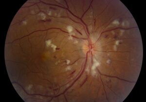
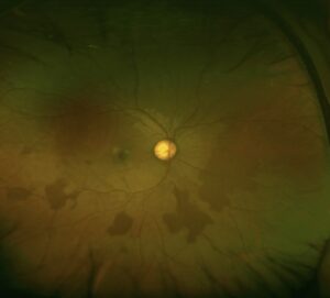
On indirect ophthalmoscopy, the retina near the ora looks finely bubbled and slightly thickened.
The cysts often appear as a grayish-white band with a stippled surface.
Next, scleral depression brings the area into profile and shows the small elevated vesicles more clearly.
Temporal quadrants usually show the most obvious changes.
How to Examine the Periphery
First, dilate fully and examine both eyes with the binocular indirect ophthalmoscope.
Then, use scleral depression across all quadrants, moving slowly from the ora backward.
A contact lens at the slit lamp can also clarify surface detail and suspicious edges.
Imaging in the Periphery
Widefield photographs can document the location, although the far periphery may remain poorly resolved.
When OCT can reach the area, it shows small intraretinal cystic spaces.
A normal central scan, however, says nothing about the far periphery.
Cystoid Degeneration vs Retinoschisis
Typical degenerative retinoschisis is thought to develop when peripheral cystoid cavities coalesce and enlarge.
Therefore, the two findings can sit next to each other in the same eye.
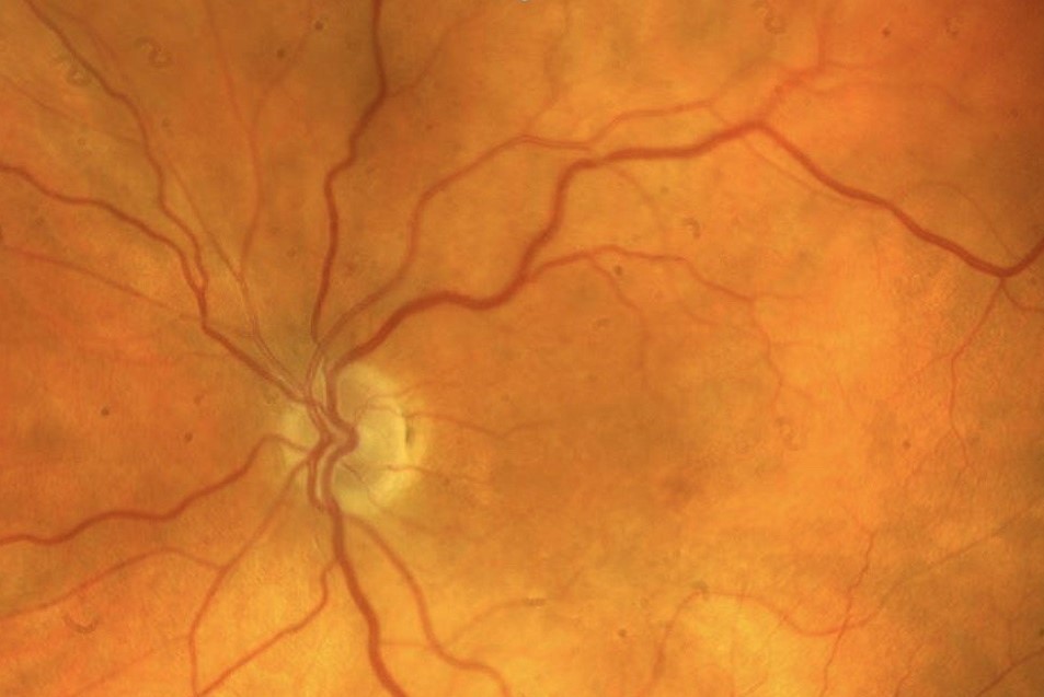
Schisis forms a smooth, fixed, dome-shaped elevation, often inferotemporally and bilaterally.
Its inner wall may show white flecks, and its vessels may look sclerosed.
An absolute field defect also favors schisis, because the split interrupts retinal signal transmission.
For a detailed comparison with fluid-filled elevations, review retinoschisis vs retinal detachment.
Cystoid Degeneration vs Retinal Breaks
Small round cysts may mimic atrophic holes, particularly when the view is limited.
A true break usually shows a sharply defined red defect, and depression can reveal its edges.
Meanwhile, a horseshoe tear has a flap and often has vitreous traction attached to it.
Cysts lack these features and typically occur as a uniform field of many small spaces.
If doubt persists, use contact lens biomicroscopy or arrange a retina review.
Differential Diagnosis of Cystoid Degeneration
Several peripheral lesions can look similar, because the view is often limited.
For example, lattice degeneration shows circumferential thinning, white lines, and pigment changes.
Also, white without pressure lacks the clustered vesicles seen in cysts.
Therefore, identify each lesion separately before you decide on follow-up.
Symptoms That Change the Plan
Isolated cystoid changes should not explain new flashes, floaters, or a curtain.
Therefore, treat those symptoms as a possible posterior vitreous detachment or tear until examination proves otherwise.
Also, when a detachment is present, use its configuration to guide the search with the Lincoff rules.
Management of Cystoid Degeneration
Asymptomatic isolated cysts need no laser, cryotherapy, or surgery.
No evidence supports prophylactic treatment for this finding, and treatment would add avoidable risk.
Instead, record the location and extent, then continue routine dilated examinations.
Management changes only when the examination reveals another lesion with its own indication (see peripheral retinal degenerations).
Follow-Up of Cystoid Degeneration
Routine dilated examination at the usual interval is enough for most patients.
However, shorten the interval in high myopia, with a family history of detachment, or after retinal surgery.
Then, document the extent with a drawing or widefield photograph.
As a result, later changes are easier to recognize.
Counseling the Patient
Explain that this is a common aging change and does not usually threaten vision.
Also, make clear that the finding does not protect against a future retinal tear or detachment.
Therefore, ask patients to report new floaters, flashes, or a shadow promptly.
Pitfalls to Avoid
- Do not assume every peripheral red spot is a cyst
- Avoid labeling a smooth bullous elevation as simple cystoid change
- Never stop the search after finding one benign lesion
- Do not treat isolated cysts with prophylactic laser
- Avoid dismissing new flashes because old peripheral changes are present


Document what you see
Two smartphone imaging tools built for everyday clinic use — one for the slit lamp, one for the fundus.
From Choroida — the team behind this siteReferences
- O’Malley PF, Allen RA. Peripheral cystoid degeneration of the retina: incidence and distribution in 1,000 autopsy eyes. Arch Ophthalmol. 1967;77:769-776.
- Foos RY, Feman SS. Reticular cystoid degeneration of the peripheral retina. Am J Ophthalmol. 1970;69:392-403.
- Straatsma BR, Foos RY. Typical and reticular degenerative retinoschisis. Am J Ophthalmol. 1973;75:551-575.
- Byer NE. Long-term natural history of senile retinoschisis with implications for management. Ophthalmology. 1986;93:1127-1137.
- Flaxel CJ, Adelman RA, Bailey ST, et al. Posterior vitreous detachment, retinal breaks, and lattice degeneration Preferred Practice Pattern. Ophthalmology. 2020;127:P146-P181.

Writes and reviews the clinical articles on Educate. Every post is checked against peer-reviewed sources before it goes live.