Optic nerve avulsion is a traumatic disinsertion of the optic nerve fibers from the globe at the lamina cribrosa.
It usually causes sudden, severe and permanent vision loss.
Because vitreous hemorrhage often hides the disc, ultrasound may provide the first evidence.
What Is Optic Nerve Avulsion?
The nerve fibers separate from the eye at the level of the disc.
However, the dural sheath often remains in continuity, so the nerve may look intact on orbital imaging.
The injury may involve part of the disc or its full circumference.
Mechanism of Optic Nerve Avulsion
Several forces may act together during the injury.
First, extreme rotation of the globe can stretch the nerve.
Next, forward displacement of the eye can add traction at the scleral canal.
In addition, a sudden rise in intraocular pressure may push the fibers backward through the lamina.
Because the lamina cribrosa is a relatively weak point, the fibers can tear while the sclera stays closed.
Fundus Explorer Pro
Photograph the retinal findings described here with the phone already in your pocket — 22 D optics and built-in illumination in one handheld unit.
From Choroida — the team behind this siteSymptoms of Optic Nerve Avulsion
Patients typically report immediate vision loss at the time of injury.
Vision may range from reduced acuity in partial cases to no light perception in complete cases.
Pupil Findings
A relative afferent pupillary defect is the expected finding in unilateral injury.
In a complete avulsion, the direct light reflex is absent while the consensual reflex persists.
However, traumatic mydriasis can complicate this examination (see sphincter injury).
Fundus Signs in Optic Nerve Avulsion
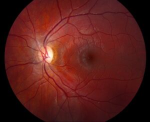
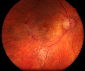
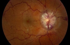
When the view allows, the examiner may see a deep pit or cavity at the expected disc position.
Hemorrhage often surrounds or fills this defect.
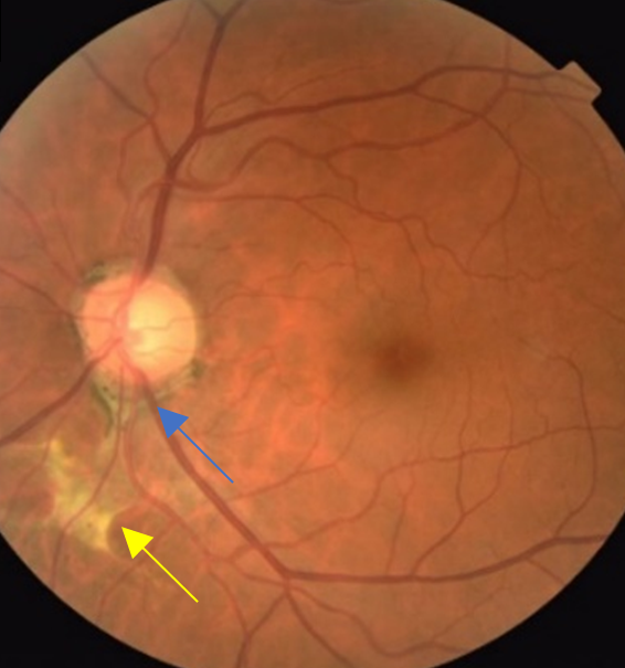
Also, a partial injury may show a crescentic excavation along one sector of the disc margin.
Retinal whitening and vascular attenuation may coexist, because the injury can also disrupt the central retinal vessels.
Associated Ocular Injuries
Always examine the whole globe, because blunt or penetrating force rarely affects one structure alone.
Look for hyphema, angle recession, commotio retinae, choroidal rupture and retinal tears.
In addition, severe orbital contact can produce disruption resembling retinitis sclopetaria.
B-Scan in Optic Nerve Avulsion
Ultrasound becomes especially useful when vitreous hemorrhage blocks the fundus view.
The scan may show a defect or hypoechoic area at the optic nerve head.
However, perform B-scan only after an open globe has been excluded or with very careful technique.
CT and MRI Findings
CT helps identify orbital fractures, foreign bodies and globe injury.
However, a normal nerve contour on CT does not exclude avulsion, because the sheath may stay intact.
MRI can add soft tissue detail, but first exclude a metallic foreign body.
Differential Diagnosis of Optic Nerve Avulsion
The history and exam help separate avulsion from other causes of post-traumatic vision loss:
- Traumatic optic neuropathy, which often shows a normal disc at first
- Posterior scleral rupture with vitreous hemorrhage and low pressure
- Central retinal artery occlusion after orbital trauma
- Congenital disc excavation, such as coloboma or an optic pit
- Choroidal rupture or sclopetaria with macular involvement
Treatment of Optic Nerve Avulsion
No treatment has proven able to reattach the torn fibers or restore lost vision.
Evidence for steroids and nerve decompression comes from traumatic optic neuropathy, and it does not establish a clear benefit.
In addition, high-dose steroids carry systemic risks, particularly after significant head injury.
Vitrectomy may clear persistent hemorrhage, but it cannot repair the nerve.
Therefore, management focuses on associated injuries, complications and protection of the fellow eye.
Visual Outlook After Optic Nerve Avulsion
Prognosis is generally poor, and complete avulsion rarely leaves useful vision.
Partial cases have a more variable course, although some retained vision may persist.
Avoid exact predictions until the fundus can be seen clearly.
Follow-Up and Late Changes
Hemorrhage gradually clears over time, then the excavation may become easier to assess.
Later, glial tissue may fill the defect while the surrounding fundus shows atrophic changes.
Meanwhile, monitor for retinal detachment, pressure rise and other delayed trauma complications.
Counseling the Patient
Explain that the nerve fibers carry visual signals and cannot currently be reconnected.
Then discuss the difference between treating complications and restoring nerve function.
Finally, advise polycarbonate protection during sports and hazardous work to protect the fellow eye.
Pitfalls to Avoid
- Do not attribute all vision loss to vitreous hemorrhage without assessing the pupil
- Never apply a B-scan probe firmly when an open globe remains possible
- Do not rule out avulsion because CT shows a continuous nerve
- Avoid promising recovery after vitrectomy or steroids


Document what you see
Two smartphone imaging tools built for everyday clinic use — one for the slit lamp, one for the fundus.
From Choroida — the team behind this siteReferences
- Foster BS, March GA, Lucarelli MJ, Samiy N, Albert DM. Optic nerve avulsion. Arch Ophthalmol. 1997;115:623-630.
- Sawhney R, Kochhar S, Gupta R, Jain R, Sood S. Traumatic optic nerve avulsion: role of ultrasonography. Eye. 2003;17:667-670.
- Steinsapir KD, Goldberg RA. Traumatic optic neuropathy. Surv Ophthalmol. 1994;38:487-518.
- Levin LA, Beck RW, Joseph MP, Seiff S, Kraker R. The treatment of traumatic optic neuropathy: the International Optic Nerve Trauma Study. Ophthalmology. 1999;106:1268-1277.

Writes and reviews the clinical articles on Educate. Every post is checked against peer-reviewed sources before it goes live.