Retinal laser recovery is usually quick, and most patients resume ordinary routines within a day.
However, the eye needs time to build a firm scar around the treated break.
Therefore, counseling should cover expected symptoms, adhesion timing, activity and warning signs.
What Happens After the Laser
Laser burns create a controlled thermal injury in the outer retina and retinal pigment epithelium.
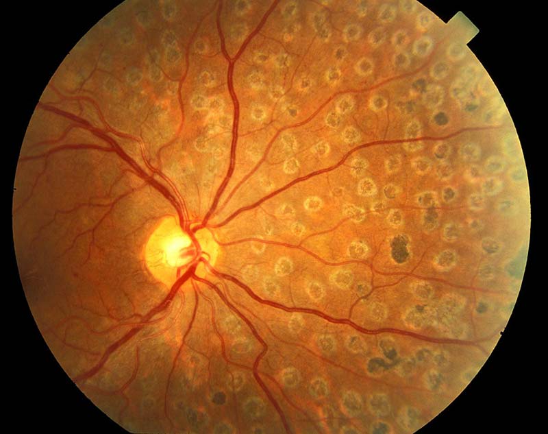
As healing progresses, a chorioretinal scar welds the retina around the break.
The tear itself stays open, because the laser only walls it off (see laser retinopexy).
As a result, fluid has a harder time spreading beyond the treated margin.
First Hours of Retinal Laser Recovery
Dilation and the bright treatment light commonly cause glare, dazzle and blurred near vision for several hours.
Coupling gel can also leave the eye sticky and the vision smeared.
Some patients feel a dull ache, and simple oral analgesia usually settles it.
Because dilation impairs focus, patients should arrange a driver for the trip home.
Fundus Explorer Pro
Photograph the retinal findings described here with the phone already in your pocket — 22 D optics and built-in illumination in one handheld unit.
From Choroida — the team behind this siteThe First Few Days
Mild irritation may follow contact lens use, and a small epithelial abrasion occasionally explains it.
Then, the glare and ache generally fade within a day or two.
Most patients need no routine drops after uncomplicated peripheral treatment, although local practice varies.
Floaters and Flashes After Treatment
Laser does not clear floaters or stop a posterior vitreous detachment.
Therefore, patients with a fresh tear may keep seeing floaters and occasional flashes for weeks.
Vitreous hemorrhage also takes time to clear, and its speed varies widely.
However, a sudden increase in symptoms needs reassessment, because it may signal a new tear (see retinal tear symptoms).
Vision During Retinal Laser Recovery
Treatment aims to lower detachment risk, so it does not improve vision.
Peripheral burns rarely produce a field defect that patients notice.
Also, preexisting floaters or hemorrhage may keep vision hazy after the procedure.
Adhesion Timing in Retinal Laser Recovery
Experimental work shows that laser increases retinal adhesion within about a day.
Yoon and Marmor, and later Folk and colleagues, documented this early effect in animal eyes.
Scar maturation then continues, and clinicians commonly quote about 10 to 14 days for maximal strength.
However, human timing data are limited, so treat this figure as an estimate.
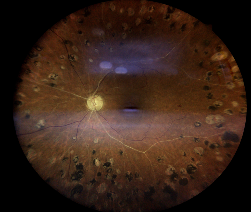
How the Scar Looks at Follow-Up
Fresh burns appear gray-white on indirect ophthalmoscopy.
Over the next one to two weeks, they develop pigmentation and a more defined border.
Next, the examiner checks for confluent rows around the whole break, including its anterior edge.
Any gap, persistent traction or fluid beyond the burns may prompt additional treatment.
Activity Advice in Retinal Laser Recovery
No strong trials define activity limits after laser for a retinal break.
Therefore, advice reflects clinician judgment and differs between practices.
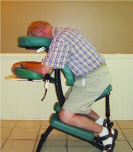
Many clinicians suggest avoiding heavy lifting, contact sports and jarring exercise for about one to two weeks.
Walking, reading, screen use and ordinary housework are generally acceptable.
Warning Signs in Retinal Laser Recovery
Patients should call the same day for any of these symptoms:
- A shower of new floaters
- New or increasing flashes
- A curtain or shadow in the side vision
- Sudden blur or distortion
- Severe pain or marked redness
A growing shadow needs urgent assessment (see retinal detachment symptoms).
Follow-Up After the Procedure
The AAO Preferred Practice Pattern suggests review one to two weeks after treating an acute symptomatic tear.
Further visits commonly follow at 4 to 6 weeks and 3 to 6 months, then annually.
Each visit should include a dilated peripheral examination of both eyes, with scleral depression when tolerated.
When Laser Fails
Detachment can still occur after well-applied laser.
For example, fluid may cross an incomplete barrier, or ongoing vitreous traction may open a new break.
Therefore, a sealed tear does not remove risk elsewhere in that eye.
Management may then involve more laser, cryotherapy or detachment surgery, depending on the findings.
Retinal Laser Recovery and Long-Term Risk
The fellow eye also deserves attention, particularly with myopia, lattice degeneration or previous detachment.
Late complications such as epiretinal membrane can occur, although they appear uncommon.
Finally, symptom advice has no expiry date, since new breaks can appear years later.
Counseling on Retinal Laser Recovery
A simple explanation works well: the laser builds a fence around the tear, and the fence strengthens over time.
Then, state clearly that treatment lowers risk without removing it.
In addition, written warning signs and a direct contact number can reduce delays in seeking care.
Pitfalls to Avoid
- Promising that floaters will disappear after treatment
- Describing the break as fully healed on the same day
- Attributing every new symptom to expected recovery
- Confusing laser advice with gas bubble restrictions
- Skipping the fellow eye at follow-up


Document what you see
Two smartphone imaging tools built for everyday clinic use — one for the slit lamp, one for the fundus.
From Choroida — the team behind this siteReferences
- Flaxel CJ, Adelman RA, Bailey ST, et al. Posterior Vitreous Detachment, Retinal Breaks, and Lattice Degeneration Preferred Practice Pattern. Ophthalmology. 2020;127:P146-P181.
- Yoon YH, Marmor MF. Rapid enhancement of retinal adhesion by laser photocoagulation. Ophthalmology. 1988;95:1385-1388.
- Folk JC, Sneed SR, Folberg R, et al. Early retinal adhesion from laser photocoagulation. Ophthalmology. 1989;96:1523-1525.
- Blindbaek S, Grauslund J. Prophylactic treatment of retinal breaks: a systematic review. Acta Ophthalmol. 2015;93:3-8.
- Hollands H, Johnson D, Brox AC, et al. Acute-onset floaters and flashes: is this patient at risk for retinal detachment? JAMA. 2009;302:2243-2249.

Writes and reviews the clinical articles on Educate. Every post is checked against peer-reviewed sources before it goes live.