The 4-2-1 rule identifies severe nonproliferative diabetic retinopathy (NPDR) on fundus examination or photographs.
It comes from Early Treatment Diabetic Retinopathy Study (ETDRS) grading, and because one positive criterion is enough, a single careful examination can change follow-up.
What the 4-2-1 Rule Means
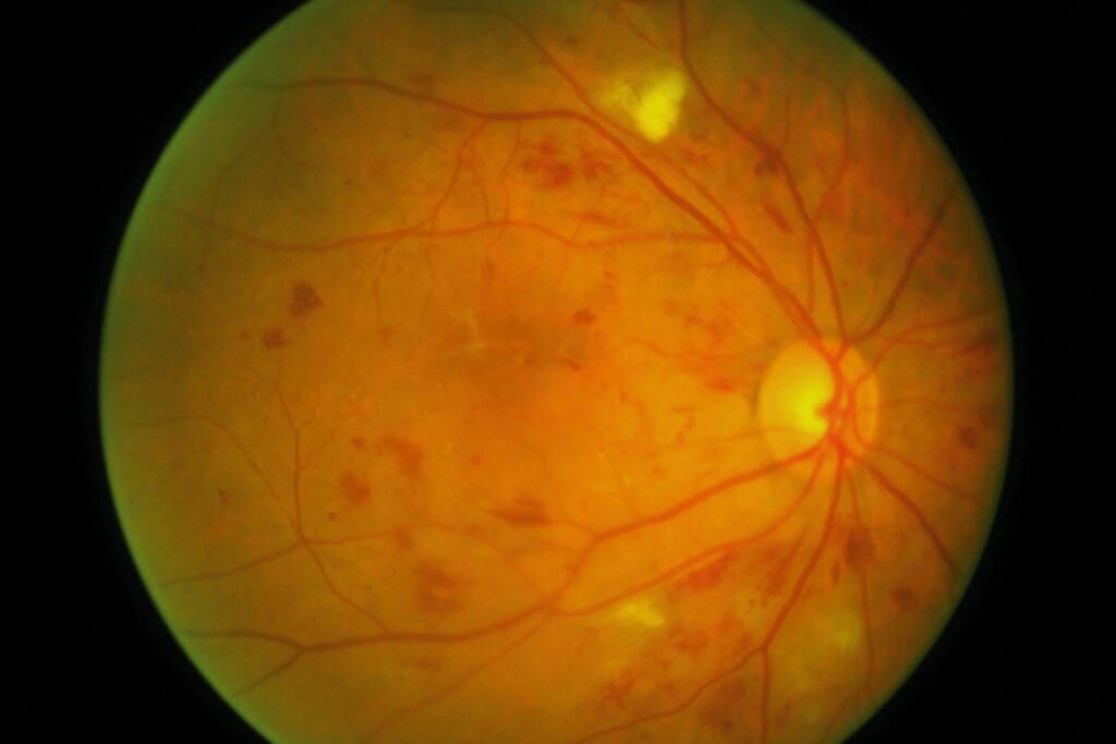
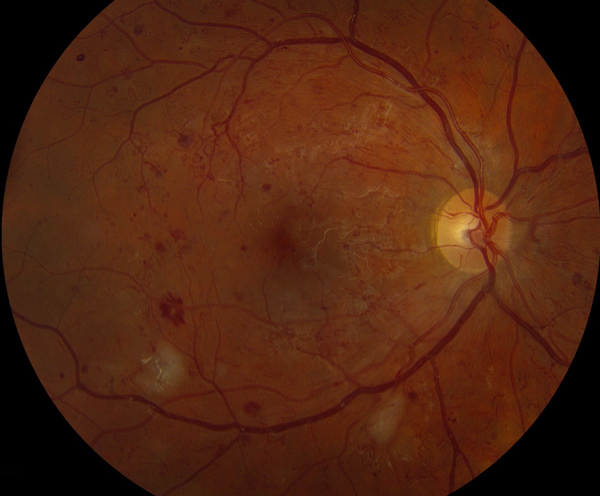
First, the 4 means severe intraretinal hemorrhages and microaneurysms in all four quadrants, while the 2 means definite venous beading in at least two quadrants. Finally, the 1 means prominent intraretinal microvascular abnormalities (IRMA) in at least one quadrant, and the eye must also show no signs of proliferative disease.
Why These Lesions Signal Ischemia
Hemorrhages and microaneurysms reflect damaged capillaries, while venous beading and IRMA often accompany nearby capillary nonperfusion. Therefore, all three suggest advancing ischemia before new vessels appear.
Fundus Explorer Pro
Photograph the retinal findings described here with the phone already in your pocket — 22 D optics and built-in illumination in one handheld unit.
From Choroida — the team behind this siteApplying the 4-2-1 Rule Step by Step
Like the Lincoff rules, this memory aid works best within a fixed routine.
- Dilate the pupil, then exclude neovascularization and preretinal or vitreous hemorrhage.
- Next, grade hemorrhages and microaneurysms in each quadrant.
- Then trace every major vein for beading.
- Search for IRMA, especially near cotton wool spots.
- Finally, record each positive criterion and its quadrants.
The 4: Hemorrhages and Microaneurysms
The ETDRS anchored this criterion to standard photograph 2A. However, the International Clinical scale offers a simpler bedside threshold: more than 20 intraretinal hemorrhages in each quadrant, and dense disease in only three quadrants therefore fails this criterion.
The 2: Venous Beading
Venous beading shows localized alternating dilation and narrowing, like a string of sausages, and the ETDRS reference is standard photograph 6B. Follow each vein outward from the disc, because subtle segments are easy to miss.
The 1: IRMA
IRMA are thin, irregular intraretinal vessels, often near cotton wool spots, and the ETDRS reference is standard photograph 8A. Because they are fine and low in contrast, a quick ophthalmoscopic look can miss them, and a large aberrant vessel crossing the macula suggests a retinal macrovessel, so it does not count.
IRMA or New Vessels?
IRMA stay within the retina and usually do not cross major vessels, and on fluorescein angiography, they typically leak little, while new vessels leak profusely. OCT can also help, because neovascularization breaches the internal limiting membrane.
Severe or Very Severe Under the 4-2-1 Rule
One positive criterion defines severe NPDR, while two or more define very severe NPDR in ETDRS terms, although the International Clinical scale has no separate very severe category. In commonly cited ETDRS estimates, about 15% of severe eyes reached high-risk proliferative disease within one year, while for very severe eyes, the figure was about 45%.
Imaging and the 4-2-1 Rule
The ETDRS based its grading on standardized seven-field stereoscopic photographs, and consequently, clinical examination can underestimate fine lesions. Angiography can also reveal nonperfusion, although the rule itself requires no angiogram.
Macular Edema Needs a Separate Check
The grade does not tell you whether macular edema is present. Therefore, examine the macula and obtain OCT at the same visit, and then report retinopathy severity and edema status separately.
Follow-Up After a Positive 4-2-1 Rule
AAO guidance supports review about every 3 to 4 months for severe NPDR without macular edema, while center-involving edema generally needs review about monthly. Also consider shorter intervals during pregnancy or after rapid glycemic improvement, and at each visit, repeat all three criteria and look again for new vessels.
Panretinal Photocoagulation
Panretinal photocoagulation (PRP) treats ischemic peripheral retina and lowers the drive for neovascularization. ETDRS Report 9 supports considering scatter treatment as eyes approach high-risk disease. However, PRP can reduce peripheral and night vision, and it can worsen macular edema, while it also cannot restore lost vision.
Anti-VEGF Therapy Before Proliferative Disease
PANORAMA studied aflibercept in moderately severe to severe NPDR without center-involving edema, and treated eyes improved in severity grade and developed fewer vision-threatening complications. Protocol W also found fewer complications, although 4-year visual acuity did not differ meaningfully from sham. Therefore, discuss injection burden, endophthalmitis risk and the continued need for monitoring.
Discussing the 4-2-1 Rule With Patients
Patients with good vision may doubt a severe label, so explain that it describes retinal vascular damage and future risk, even when sight is normal. Then review warning symptoms, such as new floaters or sudden blur.
Pitfalls to Avoid
- Do not pool quadrants across criteria, because each has its own threshold
- Never finalize the grade through an undilated pupil
- Do not call a vessel IRMA before excluding neovascularization
- Do not let good vision lower your suspicion


Document what you see
Two smartphone imaging tools built for everyday clinic use — one for the slit lamp, one for the fundus.
From Choroida — the team behind this siteReferences
- Early Treatment Diabetic Retinopathy Study Research Group. Fundus photographic risk factors for progression of diabetic retinopathy. ETDRS report number 12. Ophthalmology. 1991;98(5 Suppl):823-833.
- Early Treatment Diabetic Retinopathy Study Research Group. Early photocoagulation for diabetic retinopathy. ETDRS report number 9. Ophthalmology. 1991;98(5 Suppl):766-785.
- Wilkinson CP, Ferris FL, Klein RE, et al. Proposed international clinical diabetic retinopathy and diabetic macular edema disease severity scales. Ophthalmology. 2003;110:1677-1682.
- Flaxel CJ, Adelman RA, Bailey ST, et al. Diabetic Retinopathy Preferred Practice Pattern. Ophthalmology. 2020;127:P66-P145.
- Brown DM, Wykoff CC, Boyer D, et al. Evaluation of intravitreal aflibercept for the treatment of severe nonproliferative diabetic retinopathy: results from the PANORAMA randomized clinical trial. JAMA Ophthalmol. 2021;139:946-955.
- Maturi RK, Glassman AR, Josic K, et al. Four-year visual outcomes in the Protocol W randomized trial of intravitreal aflibercept for prevention of vision-threatening complications of diabetic retinopathy. JAMA. 2023;329:376-385.

Writes and reviews the clinical articles on Educate. Every post is checked against peer-reviewed sources before it goes live.