Pituitary apoplexy is sudden hemorrhage or infarction of the pituitary gland, usually within an unrecognized adenoma.
Patients typically present with severe headache and abrupt visual symptoms, and because the eye findings may dominate, the ophthalmologist is often the first to suspect it.
What Happens in Pituitary Apoplexy
An adenoma outgrows its blood supply, or a vessel within it bleeds, and because the bony sella cannot expand, pressure rises within hours. As a result, the swollen gland pushes up on the chiasm and sideways into the cavernous sinuses. Most patients did not know they had a tumor before the event.
Who Is at Risk
- Pituitary macroadenoma, known or undiagnosed
- Systemic hypertension
- Anticoagulant or antiplatelet therapy
- Major surgery, especially cardiac bypass
- Dopamine agonist treatment for prolactinoma
- Dynamic pituitary function testing
- Pregnancy and head trauma
All-fit Slit-Lamp Adapter
Record and share exactly what you see at the slit lamp. One adapter fits any slit lamp or surgical microscope — and any smartphone.
From Choroida — the team behind this sitePresenting Symptoms
Headache is the most common complaint, and it is usually sudden, severe and retro-orbital (see headache red flags). Nausea, vomiting and neck stiffness may follow, and then the patient notices blurred vision, a missing half of the field or double vision. Some patients also become drowsy or confused, which makes the visual history unreliable.
Visual Acuity in Pituitary Apoplexy
Acuity can range from normal to no light perception, but loss is often asymmetric because the mass compresses the chiasm unevenly. Therefore, test each eye separately with best correction, and also check color vision, since it may fall before acuity does.
Visual Field Defects
Bitemporal hemianopia is the classic pattern of chiasmal compression, but apoplexy often produces irregular, asymmetric or monocular defects. For example, a junctional scotoma combines central loss in one eye with a temporal defect in the other. Confrontation testing at the bedside still helps when the patient cannot sit for perimetry.
Ocular Motor Palsies in Pituitary Apoplexy
Lateral expansion compresses the cranial nerves within the cavernous sinus. The third nerve suffers most often, causing ptosis, a dilated pupil and limited adduction, while sixth and fourth nerve palsies follow in frequency. However, sudden headache with a pupil-involving third nerve palsy also suggests a posterior communicating artery aneurysm.
Pupils and the Optic Disc
Look for a relative afferent pupillary defect when compression is asymmetric, although a coexisting third nerve palsy can make the swinging flashlight test difficult to read. In the acute phase, the optic discs usually look normal, but pallor appears weeks later, so a normal disc never excludes compression.
Differential Diagnosis of Pituitary Apoplexy
Several emergencies share sudden headache with visual loss (see sudden vision loss).
- Subarachnoid hemorrhage from a ruptured aneurysm
- Bacterial meningitis
- Cavernous sinus thrombosis
- Migraine with aura or ophthalmoplegia
- Hypophysitis or pituitary metastasis
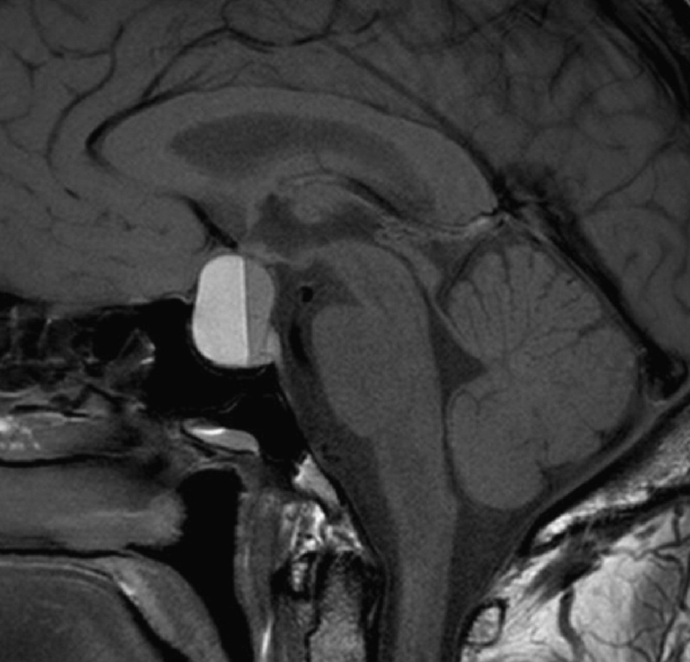
Imaging for Pituitary Apoplexy
MRI is the preferred study, and it shows a sellar mass with hemorrhage or necrosis. Emergency teams often order CT first because it is fast, but CT may miss a small bleed within the adenoma, so add vascular imaging when an aneurysm is still possible.
The Endocrine Emergency
Acute corticotropin deficiency is the main threat to life, so draw blood for cortisol and other pituitary hormones, then start intravenous hydrocortisone without waiting for results. Sodium and fluid balance also need close monitoring, and finally, never send the patient home to await an outpatient scan.
Surgery in Pituitary Apoplexy
Transsphenoidal decompression removes the hemorrhagic tumor and relieves pressure on the chiasm, and the UK guidelines favor early surgery for severe acuity loss, dense field defects or deteriorating vision. Surgery cannot restore optic nerve fibers that have already died, and in addition, it carries risks such as CSF leak, diabetes insipidus and new hormone deficiency.
Conservative Management
Many centers manage isolated ocular palsies or mild, stable field loss without surgery, and these patients need steroid cover and daily checks of acuity and fields. Any decline in vision should then prompt urgent neurosurgical review, while retrospective series suggest similar outcomes in carefully selected patients.
Visual Outcome After Pituitary Apoplexy
Ocular motor palsies usually recover over weeks to months with either approach, and acuity and fields also improve in many patients after decompression. However, severe initial loss and established optic atrophy predict a poorer result, and some series favor early surgery, although selection bias limits these data.
Counseling the Patient
Explain that the bleed occurred inside a pituitary tumor that is usually benign, and that double vision often resolves, although recovery can take several months. Next, discuss hormone replacement, because many patients need it long term, and also advise against driving until formal fields confirm a safe result.
Follow-Up Eye Examination
Repeat acuity, color vision and formal perimetry after treatment, while OCT of the ganglion cell layer may also help, since thinning suggests limited recovery potential. Later MRI scans check for tumor regrowth, and persistent diplopia may need prisms or, occasionally, strabismus surgery.
Pitfalls to Avoid
- Do not label sudden headache with diplopia as migraine
- Never delay steroids while waiting for imaging
- Do not trust a normal disc to exclude compression
- Avoid testing visual fields only binocularly


Document what you see
Two smartphone imaging tools built for everyday clinic use — one for the slit lamp, one for the fundus.
From Choroida — the team behind this siteReferences
- Rajasekaran S, Vanderpump M, Baldeweg S, et al. UK guidelines for the management of pituitary apoplexy. Clin Endocrinol (Oxf). 2011;74:9-20.
- Briet C, Salenave S, Bonneville JF, Laws ER, Chanson P. Pituitary apoplexy. Endocr Rev. 2015;36:622-645.
- Randeva HS, Schoebel J, Byrne J, Esiri M, Adams CB, Wass JA. Classical pituitary apoplexy: clinical features, management and outcome. Clin Endocrinol (Oxf). 1999;51:181-188.
- Ayuk J, McGregor EJ, Mitchell RD, Gittoes NJ. Acute management of pituitary apoplexy: surgery or conservative management? Clin Endocrinol (Oxf). 2004;61:747-752.

Writes and reviews the clinical articles on Educate. Every post is checked against peer-reviewed sources before it goes live.