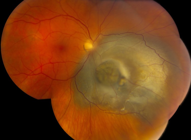
Collar button melanoma describes a choroidal melanoma that has broken through Bruch’s membrane and taken a mushroom shape.
Few other choroidal masses adopt this profile, so the sign has real diagnostic value.
However, the shape alone does not establish the diagnosis or set the treatment.
What Collar Button Melanoma Means
The tumor has a broad choroidal base, a narrow waist and a rounded subretinal head.
Therefore, its profile resembles a shirt collar stud or a mushroom.
Most uveal melanomas remain dome-shaped.
How Bruch’s Membrane Creates the Shape
Early growth occurs beneath Bruch’s membrane, which initially restrains the tumor.
Then, continued pressure can rupture the membrane and overlying retinal pigment epithelium.
As a result, tumor cells herniate into the subretinal space.
The rigid rim of the break forms the waist, while the head expands freely.
Fundus Explorer Pro
Photograph the retinal findings described here with the phone already in your pocket — 22 D optics and built-in illumination in one handheld unit.
From Choroida — the team behind this siteSymptoms of Collar Button Melanoma
Patients may report blurred vision, floaters, photopsia or a field defect.
Some have no symptoms, especially when the tumor lies away from the macula.
Also, vitreous hemorrhage can present as sudden visual loss and hide the mass.
Therefore, unexplained hemorrhage in an adult requires ultrasound when the fundus view is poor.
Fundus Signs to Look For
Indirect ophthalmoscopy shows an elevated mass with a visible shoulder near the base.
Color varies from dark brown to amelanotic.
Also look for orange pigment, subretinal fluid, surface hemorrhage and dilated vessels.
Because the head can overhang the base, the fundus view may underestimate the footprint.
Collar Button Melanoma on B-Scan Ultrasound
B-scan offers the clearest demonstration of the shape, even through media opacity.
First, scan the tumor center in two perpendicular planes, because an oblique section can hide the waist.
Next, look for a broad base, a constricted neck and an expanded subretinal head.
Acoustic hollowness and choroidal excavation support the diagnosis.
A-Scan, Angiography and OCT
A-scan typically shows low to medium internal reflectivity with a regular structure.
However, hemorrhage and necrosis can alter these patterns.
Fluorescein angiography may show intrinsic tumor vessels and late leakage.
OCT helps assess macular fluid, but its limited depth rarely captures a large mass.
Therefore, ultrasound remains the main tool for shape.
Differential in Collar Button Melanoma
The exam should explain every feature, since other lesions can look similar:
- Choroidal metastasis, usually with a known primary
- Choroidal melanocytoma with focal necrosis
- Peripheral exudative hemorrhagic chorioretinopathy
- Suprachoroidal hemorrhage, which evolves over serial scans
- Amelanotic hemangioma or lymphoid lesions
Measuring the Tumor Accurately
Measure thickness from the inner sclera to the tumor apex.
Also record the largest basal diameter and the distances to the disc and fovea.
Serial scans should use the same technique, because oblique sections can create false growth.
Staging and Prognosis in Collar Button Melanoma
Current AJCC staging relies on thickness, basal diameter, ciliary body involvement and extraocular extension.
Therefore, the mushroom shape does not create a separate stage.
Metastatic risk rises with tumor thickness, and mushroom tumors are often large at diagnosis.
Genetic Prognostic Testing
Gene expression profiling can stratify metastatic risk in uveal melanoma.
However, biopsy carries sampling limits and procedural risks, including hemorrhage.
Therefore, discuss testing as a prognostic tool, because it does not treat the tumor.
Treatment Choices for Collar Button Melanoma
Treatment depends on tumor size, location, visual potential and systemic status (see choroidal melanoma overview).
Options include plaque brachytherapy, proton beam radiotherapy and enucleation.
For medium tumors, COMS found no significant mortality difference between iodine-125 brachytherapy and enucleation.
However, very large tumors may not suit eye-sparing treatment, so refer early to an ocular oncology center.
Limits and Risks of Treatment
Radiation aims for local control, although useful vision may still decline.
Radiation retinopathy, optic neuropathy, cataract and neovascular glaucoma can follow treatment.
In addition, local control cannot guarantee protection from metastasis.
Counseling Patients With Collar Button Melanoma
Explain that the break lies inside the eye, between the choroid and the retina.
Thus, the sign does not mean that cancer has left the eye.
Next, separate three goals: tumor control, vision and systemic surveillance.
Because vision outcomes vary, avoid firm predictions early.
Follow-Up After Treatment
Regular examination and ultrasound track tumor height, reflectivity and marginal recurrence.
A treated tumor may keep some mushroom profile, so stable shape does not indicate failure.
Meanwhile, examine for radiation complications at each visit.
Systemic surveillance, often including liver imaging, follows a schedule set by risk.
Pitfalls to Avoid
- Do not rule out melanoma because the tumor lacks pigment
- Avoid calling the waist absent after one oblique scan
- Never equate Bruch’s membrane rupture with extrascleral extension
- Do not delay referral while waiting to document growth


Document what you see
Two smartphone imaging tools built for everyday clinic use — one for the slit lamp, one for the fundus.
From Choroida — the team behind this siteReferences
- Shields CL, Manalac J, Das C, Ferguson K, Shields JA. Choroidal melanoma: clinical features, classification, and top 10 pseudomelanomas. Curr Opin Ophthalmol. 2014;25:177-185.
- Shields CL, Furuta M, Thangappan A, et al. Metastasis of uveal melanoma millimeter-by-millimeter in 8033 consecutive eyes. Arch Ophthalmol. 2009;127:989-998.
- Collaborative Ocular Melanoma Study Group. The COMS randomized trial of iodine 125 brachytherapy for choroidal melanoma: V. Twelve-year mortality rates and prognostic factors: COMS report No. 28. Arch Ophthalmol. 2006;124:1684-1693.
- Onken MD, Worley LA, Char DH, et al. Collaborative Ocular Oncology Group report number 1: prospective validation of a multi-gene prognostic assay in uveal melanoma. Ophthalmology. 2012;119:1596-1603.

Writes and reviews the clinical articles on Educate. Every post is checked against peer-reviewed sources before it goes live.