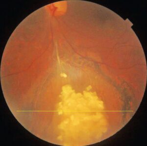
Headlight in fog describes a bright white retinal focus seen through dense vitreous haze.
Clinicians link this sign most strongly with active toxoplasma retinochoroiditis.
However, it describes an appearance, so it cannot confirm the diagnosis alone.
Therefore, interpret it alongside the lesion, any nearby scar and the patient’s immune status.
What Headlight in Fog Looks Like
The fundus view looks gray and hazy, like fog over a windshield.
Through it, a fluffy white lesion glows with indistinct borders.
Vessels and disc details may blur, while the focus still stands out.
In addition, indirect ophthalmoscopy shows the lesion best against the haze.
Why the Sign Appears
Toxoplasma destroys retinal tissue, which produces a dense white patch of necrosis.
Then inflammatory cells enter the overlying vitreous and scatter light.
However, the necrotic retina reflects light strongly, so it remains visible.
Fundus Explorer Pro
Photograph the retinal findings described here with the phone already in your pocket — 22 D optics and built-in illumination in one handheld unit.
From Choroida — the team behind this siteSymptoms With Headlight in Fog
Patients usually report floaters and blurred vision, often in one eye.
Pain and redness may also occur when anterior uveitis accompanies the focus.
Finally, haze alone can reduce acuity, so vision does not locate the lesion.
Diagnostic Weight of Headlight in Fog
The sign raises strong suspicion for toxoplasmosis, especially in an immunocompetent patient.
Its value increases when the active focus lies beside an old pigmented scar.
However, reliable sensitivity and specificity figures are lacking, so treat it as a clinical clue.
Findings That Support Toxoplasmosis
A satellite lesion next to a chorioretinal scar forms the classic pattern.
Nearby vessels may also show sheathing, and granulomatous anterior uveitis is common.
The fellow eye may hold a silent scar, so examine both eyes.
Atypical Toxoplasmosis
Immunosuppressed and older patients may develop large, multifocal or bilateral lesions.
A pigmented scar may be absent, so severe disease can resemble viral necrotizing retinitis.
Therefore, these patients often need early intraocular fluid testing.
Viral Retinitis Mimicking Headlight in Fog
Acute retinal necrosis usually begins peripherally and causes occlusive arteritis.
Its vitritis can be dense, so early cases may look similar.
CMV retinitis, by contrast, causes little vitritis in severely immunosuppressed patients.
Therefore, rapid progression or peripheral whitening should prompt urgent viral PCR.
Syphilis and Tuberculosis
Syphilis can produce necrotizing retinitis, placoid lesions and vitritis in varied patterns.
Therefore, order treponemal serology in every patient with unexplained posterior inflammation.
Tuberculosis may also present with a choroidal granuloma, subretinal abscess or vasculitis.
Next, use exposure history, interferon-gamma testing and chest imaging to assess that risk.
Endophthalmitis as a Mimic
Endogenous fungal disease causes fluffy chorioretinal lesions with overlying vitreous opacities (see ocular candidiasis).
Ask about central lines, injection drug use, recent surgery and hospital admission.
Because delay can cost vision, these patients need same-day assessment.
Lymphoma and Other Masquerades
Intraocular lymphoma typically affects older adults and may cause persistent vitreous cells.
Steroids can temporarily clear the haze, which can falsely reassure the clinician.
Therefore, consider vitreous biopsy when inflammation persists or recurs without a clear cause.
Toxocara and Behçet disease can also cause a hazy focus (see retinal vasculitis).
Workup of Headlight in Fog
Toxoplasma IgG confirms past exposure, but it cannot prove the lesion is toxoplasmic.
However, a negative IgG makes the diagnosis much less likely.
For atypical cases, aqueous PCR and the Goldmann-Witmer coefficient provide local evidence.
Imaging Through Haze
OCT may show a thickened, hyperreflective retina with disrupted layers at the active focus.
Fluorescein angiography typically shows early hypofluorescence, then late leakage and staining.
When opacities block the view, B-scan helps exclude retinal detachment.
Managing Headlight in Fog
Clinicians usually treat lesions threatening the macula or disc, large lesions and severe vitritis.
Evidence for preventing permanent damage remains modest (see treatment regimens).
Also, avoid corticosteroids without antimicrobial cover, because they can worsen necrotizing infection.
Follow-up After the Haze Clears
As inflammation settles, the border sharpens and pigmentation develops.
Meanwhile, monitor intraocular pressure, vitreous traction and choroidal neovascularization.
If the lesion enlarges despite therapy, reconsider the diagnosis.
Counseling Patients With Headlight in Fog
Explain that the name describes the examination view and does not name a separate disease.
Because scars can reactivate, patients should report new floaters promptly.
Treatment controls inflammation but cannot restore destroyed retina.
Pitfalls to Avoid
- Do not diagnose toxoplasmosis from the haze pattern alone
- Do not treat positive IgG as proof of active infection
- Never give steroids alone for an undiagnosed necrotizing focus
- Do not skip syphilis testing in posterior uveitis


Document what you see
Two smartphone imaging tools built for everyday clinic use — one for the slit lamp, one for the fundus.
From Choroida — the team behind this siteReferences
- Holland GN. Ocular toxoplasmosis: a global reassessment. Part I: epidemiology and course of disease. Am J Ophthalmol. 2003;136:973-988.
- Holland GN. Ocular toxoplasmosis: a global reassessment. Part II: disease manifestations and management. Am J Ophthalmol. 2004;137:1-17.
- Butler NJ, Furtado JM, Winthrop KL, Smith JR. Ocular toxoplasmosis II: clinical features, pathology and management. Clin Exp Ophthalmol. 2013;41:95-108.
- Park YH, Nam HW. Clinical features and treatment of ocular toxoplasmosis. Korean J Parasitol. 2013;51:393-399.
- Smith JR, Ashander LM, Arruda SL, et al. Pathogenesis of ocular toxoplasmosis. Prog Retin Eye Res. 2021;81:100882.

Writes and reviews the clinical articles on Educate. Every post is checked against peer-reviewed sources before it goes live.