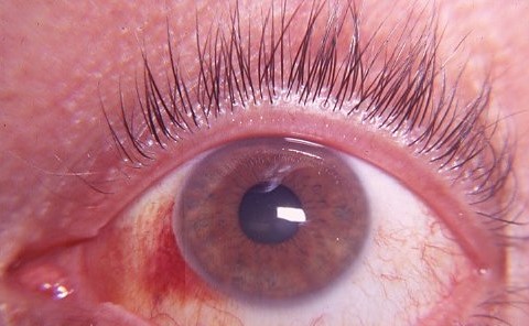
Subconjunctival hemorrhage healing usually takes one to two weeks, although large bleeds may need about three weeks.
The blood clears by gradual resorption, and no drop has a proven ability to speed it.
Therefore, the clinician should confirm the diagnosis, set expectations and identify the few cases that need workup.
Why Subconjunctival Hemorrhage Healing Takes Weeks
Blood collects beneath the conjunctiva after a small vessel ruptures.
Because this tissue plane is loose, a small volume can cover a large area.
Macrophages then break down the red cells, while local drainage clears the remnants.
Subconjunctival Hemorrhage Healing Timeline
These ranges reflect typical clinical experience, so individual cases may vary.
- Days 1 to 2: a flat, bright red patch that may still enlarge
- During days 3 to 7, the patch stays stable or fades slowly
- Then, by days 7 to 14, the edges fade with orange or yellow tones
- Finally, weeks 2 to 3 bring faint yellow residue after larger bleeds
All-fit Slit-Lamp Adapter
Record and share exactly what you see at the slit lamp. One adapter fits any slit lamp or surgical microscope — and any smartphone.
From Choroida — the team behind this siteColor Changes in Subconjunctival Hemorrhage Healing
Fresh blood usually looks bright red.
Later, hemoglobin breakdown produces orange and yellow tones, much like a fading skin bruise.
Therefore, tell patients that a yellow tint signals expected recovery.
History That Frames the Bleed
Start with onset, trauma, contact lens wear and recent eye surgery.
Next, ask about coughing, vomiting, heavy lifting and constipation.
Also review hypertension, diabetes, bleeding disorders and every blood-thinning medication.
Slit Lamp Findings
A typical bleed appears as a flat, sharply bordered red area with clear overlying conjunctiva.
Vision, pupils, pressure and the cornea remain normal in the uncomplicated case.
Check the anterior chamber, because blood there changes the diagnosis (see hyphema).
Then lift the lids and inspect the fornices for a foreign body, laceration or mass.
Differential Diagnosis During Subconjunctival Hemorrhage Healing
Several conditions can mimic a bleed, so confirm that the blood lies beneath clear conjunctiva.
- Conjunctivitis, because individual dilated vessels remain visible
- Episcleritis or scleritis, since deeper vessels and tenderness are present
- Conjunctival neoplasia, which may look like a flat bleed that never fades
- Also Kaposi sarcoma, especially in immunocompromised patients
Trauma and Subconjunctival Hemorrhage Healing
After blunt or penetrating injury, a hemorrhage can hide serious damage beneath it.
For example, extensive bullous bleeding may conceal an open globe.
Therefore, assess vision, motility and proptosis, and arrange imaging when the mechanism suggests it.
Contact Lens Wearers
Contact lens wearers often develop small bleeds from mechanical irritation.
However, always check lens fit and the corneal surface with fluorescein.
Also, advise a short break from lenses until the eye is quiet.
Therefore, treat pain or infiltrates as keratitis until proven otherwise.
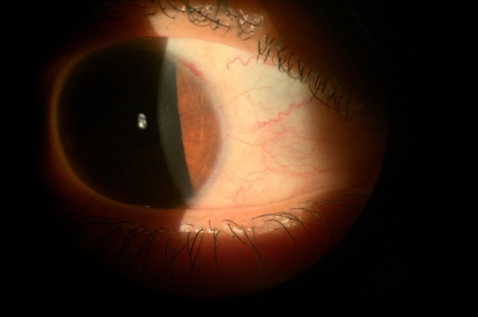
Pain and Blurred Vision Suggest Another Diagnosis
Simple bleeds cause mild irritation at most.
Significant pain, photophobia or reduced vision therefore calls for a wider search.
Consider keratitis, uveitis, scleritis and acute angle-closure glaucoma when these symptoms appear.
When Subconjunctival Hemorrhage Healing Stalls
Reassess any patch that has not clearly improved after about three weeks.
A persistent lesion may represent a conjunctival tumor, lymphangiectasia or another vascular abnormality.
Similarly, a salmon-colored or thickened area needs careful inspection and possibly biopsy.
Recurrent Bleeds Need a Different Question
One isolated episode rarely needs laboratory testing.
However, repeated episodes should prompt blood pressure measurement and a medication review.
Then consider a complete blood count and coagulation studies when history suggests a bleeding tendency.
Children and Infants
Birth trauma can cause subconjunctival bleeding in newborns, and it typically resolves without treatment.
In older children, however, an unexplained bleed deserves more caution.
Consider severe coughing, bleeding disorders and nonaccidental trauma.
Treatment During Subconjunctival Hemorrhage Healing
Observation is sufficient for the uncomplicated case.
Lubricating drops can ease mild irritation, but they do not clear the blood faster.
Likewise, antibiotic and steroid drops have no routine role.
Managing Blood Thinners
Never advise patients to stop a prescribed anticoagulant because of an isolated bleed.
The systemic risk of stopping usually outweighs a cosmetic eye finding.
Instead, contact the prescribing clinician when bleeding recurs or the INR is out of range.
Counseling About Subconjunctival Hemorrhage Healing
Show the patient the expected color sequence, because many fear that a change means worsening.
Explain that the blood sits on the eye surface and does not normally affect sight.
Advise them to return for pain, reduced vision, discharge, new trauma or no improvement by three weeks.
For more on causes and diagnosis, see the subconjunctival hemorrhage overview.
Pitfalls to Avoid
- First, do not reassure a trauma patient before excluding an open globe
- Also, avoid extensive tests after one typical painless bleed
- However, repeated bleeds in patients taking warfarin need review


Document what you see
Two smartphone imaging tools built for everyday clinic use — one for the slit lamp, one for the fundus.
From Choroida — the team behind this siteReferences
- Tarlan B, Kiratli H. Subconjunctival hemorrhage: risk factors and potential indications. Clin Ophthalmol. 2013;7:1163-1170.
- Mimura T, Usui T, Yamagami S, et al. Recent causes of subconjunctival hemorrhage. Ophthalmologica. 2010;224:133-137.
- Hu DN, Mou CH, Chao SC, et al. Incidence of non-traumatic subconjunctival hemorrhage in a nationwide study in Taiwan from 2000 to 2011. PLoS One. 2015;10:e0132762.
- Superstein R, Gomolin JE, Hammouda W, et al. Prevalence of ocular hemorrhage in patients receiving warfarin therapy. Can J Ophthalmol. 2000;35:385-389.
- American Academy of Ophthalmology. External Disease and Cornea. Basic and Clinical Science Course, Section 8. San Francisco: AAO.

Writes and reviews the clinical articles on Educate. Every post is checked against peer-reviewed sources before it goes live.