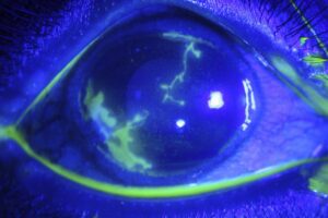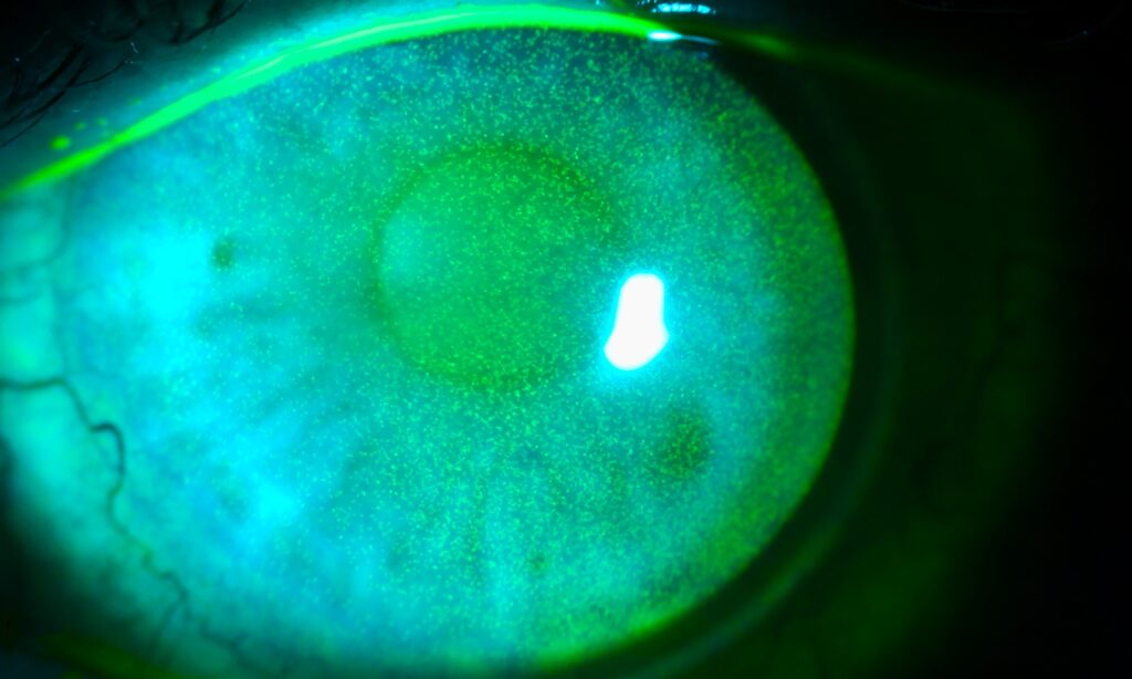
Punctate epithelial erosions are tiny, slightly depressed epithelial defects that stain with fluorescein.
They signal surface stress, although they do not identify a specific disease.
However, their position on the cornea often points toward the cause.
What Punctate Epithelial Erosions Look Like
Cobalt blue light shows fine, scattered, pinpoint green spots.
In white light, the same area may show only faint surface irregularity.
Because the lesions involve the epithelium, the underlying stroma usually stays clear.
Symptoms of Punctate Epithelial Erosions
Patients usually report grittiness, burning, photophobia or fluctuating vision.
However, symptoms and staining often correlate poorly.
For example, a neurotrophic cornea may stain heavily with little discomfort.
Therefore, test corneal sensation when the findings exceed the complaints.
All-fit Slit-Lamp Adapter
Record and share exactly what you see at the slit lamp. One adapter fits any slit lamp or surgical microscope — and any smartphone.
From Choroida — the team behind this siteHow to Map the Staining
First, instill a small amount of fluorescein and wait a minute or two.
Next, add a yellow barrier filter to the cobalt light for better contrast.
Then lift the upper lid, because superior staining hides easily.
Finally, record the location on a simple corneal drawing (see corneal staining patterns).
Inferior Punctate Epithelial Erosions
Inferior staining usually reflects lid disease or incomplete closure.
Common causes include:
- Chronic blepharitis and meibomian gland dysfunction
- Lagophthalmos, including nocturnal exposure
- Ectropion, entropion or misdirected lashes
Ask the patient to close gently, because mild lagophthalmos is easy to miss.
Interpalpebral Punctate Epithelial Erosions
A horizontal band across the open fissure suggests evaporation or ultraviolet injury.
Dry eye disease commonly produces this pattern, often with nasal and temporal conjunctival staining.
In addition, reduced blinking during screen work may worsen it.
Welding or snow exposure causes a similar band, while the history usually separates them.
Superior Punctate Epithelial Erosions
Superior staining should prompt eversion of the upper lid.
Several conditions favor this location:
- Superior limbic keratoconjunctivitis, often with thyroid disease
- Vernal keratoconjunctivitis with giant tarsal papillae
- Floppy eyelid syndrome, which often accompanies obstructive sleep apnea
- A poorly fitting contact lens
Inferonasal Staining and Drop Toxicity
Medication toxicity often concentrates inferonasally, probably because tears carry drugs toward the puncta.
Benzalkonium chloride, aminoglycosides, topical antivirals and glaucoma drops are frequent suspects.
Therefore, review every bottle, including products bought without a prescription.
Diffuse Punctate Epithelial Erosions
Widespread staining suggests infection, severe surface disease or toxicity.
Adenoviral keratoconjunctivitis typically adds follicles, watery discharge and a preauricular node.
Chlamydial disease also causes follicles, but it follows a more chronic course (see follicles versus papillae).
Next, consider severe dry eye when staining covers the whole cornea and conjunctiva.
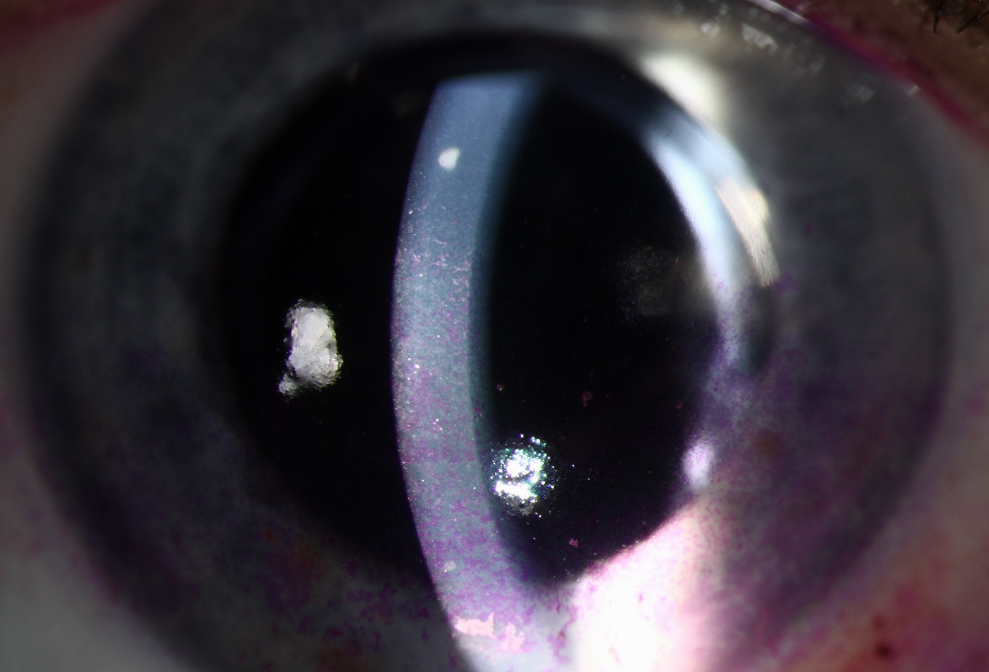
Conditions That Mimic Punctate Epithelial Erosions
Punctate epithelial keratitis shows swollen, gray-white epithelial lesions that may look slightly raised.
Thygeson keratitis, for example, produces coarse lesions in a relatively quiet eye.
Subepithelial infiltrates lie deeper and usually do not stain (see corneal infiltrates).
Also, look closely for early herpetic lesions, especially when sensation falls in one eye.
Tests That Refine the Diagnosis
Tear breakup time and Schirmer testing help assess dry eye.
In addition, lissamine green shows conjunctival damage that fluorescein may underrepresent.
Severe dryness with dry mouth should prompt evaluation for Sjögren disease.
Treating Punctate Epithelial Erosions
Treatment works best when it targets the mechanism.
Preservative-free lubricants support the surface and may ease symptoms, although responses vary between patients.
However, they cannot correct exposure, lash contact or lid malposition.
Lid hygiene, bedtime ointment, lash removal or lid surgery may therefore be needed.
When a drop causes toxicity, stop or replace it whenever the underlying disease allows.
Contact Lens Wearers With Punctate Epithelial Erosions
Contact lens wear commonly causes staining from hypoxia, solution toxicity or a poor lens fit.
Therefore, ask about wearing schedule, overnight use and the care solution.
First, pause lens wear until the staining clears, then refit with a different material or solution if needed.
Follow-Up for Punctate Epithelial Erosions
Review patients within one to two weeks after changing treatment, because the response guides the next step.
Also, repeat staining and compare it with the earlier drawing.
However, persistent staining despite appropriate therapy should prompt a second look at the diagnosis.
Counseling Patients With Punctate Epithelial Erosions
Explain that the spots are superficial and usually heal without scarring when the cause is controlled.
Also, warn that chronic drying or exposure can bring them back.
Encourage patients to report new pain, marked redness or falling vision promptly.
Those symptoms may suggest infection or a larger epithelial defect (see recurrent corneal erosion).
Pitfalls to Avoid
- Do not skip upper lid eversion
- Never label all staining as dry eye
- Avoid adding drops before reviewing current drops
- Do not overlook reduced corneal sensation


Document what you see
Two smartphone imaging tools built for everyday clinic use — one for the slit lamp, one for the fundus.
From Choroida — the team behind this siteReferences
- Bron AJ, Evans VE, Smith JA. Grading of corneal and conjunctival staining in the context of other dry eye tests. Cornea. 2003;22:640-650.
- Wolffsohn JS, Arita R, Chalmers R, et al. TFOS DEWS II diagnostic methodology report. Ocul Surf. 2017;15:539-574.
- Baudouin C, Labbé A, Liang H, et al. Preservatives in eyedrops: the good, the bad and the ugly. Prog Retin Eye Res. 2010;29:312-334.
- Bowling B. Kanski’s Clinical Ophthalmology: A Systematic Approach. 8th ed. Elsevier; 2016.

Writes and reviews the clinical articles on Educate. Every post is checked against peer-reviewed sources before it goes live.