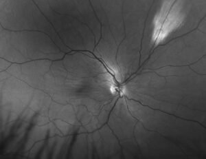
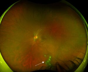
Flame hemorrhages are superficial retinal bleeds that lie within the nerve fiber layer and follow its axons.
Clinicians also call them splinter hemorrhages because of their narrow, feathered shape.
Their appearance localizes the blood, although it does not establish the cause.
Therefore, each new finding should prompt a search for a local or systemic explanation.
Anatomy of Flame Hemorrhages
Blood usually escapes from the superficial capillary network or the radial peripapillary capillaries.
It then enters the nerve fiber layer, where axons run parallel to the retinal surface.
As a result, the blood spreads along these bundles and forms a streak.
Flame Hemorrhages on Examination
Ophthalmoscopy shows bright red, linear hemorrhages with feathered ends.
Their long axis follows the nerve fiber pattern and generally points toward the disc.
They cluster around the disc and posterior pole because the nerve fiber layer is thickest there.
Fundus Explorer Pro
Photograph the retinal findings described here with the phone already in your pocket — 22 D optics and built-in illumination in one handheld unit.
From Choroida — the team behind this siteExamination Pearls
First, dilate the pupil and scan all four quadrants, because peripheral bleeds are easy to miss.
Next, compare both eyes, since asymmetry points toward occlusion.
Also, use a red-free filter, because it increases the contrast of blood against the nerve fiber layer.
Then, record the number and location of the hemorrhages for later comparison.
Flame Hemorrhages Versus Deeper Bleeds
Depth determines shape (see retinal hemorrhage patterns).
Dot and blot hemorrhages sit deeper, where vertically oriented tissue confines blood into round spots.
By contrast, preretinal blood often settles into a boat shape with a flat upper level.
Imaging Flame Hemorrhages
Color photography documents the number and distribution for later comparison.
OCT shows hyperreflective material in the inner retina, with shadowing of deeper layers.
Fluorescein angiography also reveals capillary nonperfusion or leakage that helps identify the cause.
What the Patient Reports
Many patients have no symptoms, and the finding appears during a routine examination.
However, vision may fall when macular edema, ischemia or foveal blood accompanies the hemorrhage.
Sudden unilateral blur suggests vein occlusion.
Hypertensive Flame Hemorrhages
Hypertension is a classic cause, especially when the findings are bilateral.
Examiners often also see cotton wool spots, hard exudates and arteriolar narrowing.
The Keith-Wagener-Barker system places hemorrhages and exudates in group 3.
Also, Wong and Mitchell classify these signs as moderate retinopathy, which carries a stronger association with stroke.
Retinal Vein Occlusion
Central retinal vein occlusion typically produces hemorrhages in all four quadrants, with dilated, tortuous veins.
Branch occlusion instead creates a sector of hemorrhage, often with its apex at an arteriovenous crossing.
Because both forms are usually unilateral, asymmetry helps separate them from hypertensive retinopathy.
Diabetes more typically produces dot and blot hemorrhages, but flame-shaped bleeds can appear when hypertension coexists.
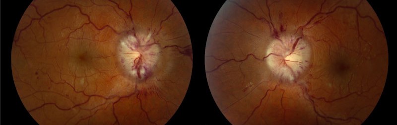
Flame Hemorrhages at the Optic Disc
A splinter hemorrhage at the disc margin has a separate differential.
In glaucoma, it may signal progression (see optic disc hemorrhage in glaucoma).
Peripapillary hemorrhages with disc swelling suggest papilledema, optic neuritis or ischemic optic neuropathy.
Blood Disorders
Severe anemia, thrombocytopenia and leukemia can all cause superficial retinal hemorrhages.
Some bleeds have white centers, known as Roth spots.
Although endocarditis is the textbook association, Roth spots also occur with leukemia and anemia.
Therefore, the finding justifies a complete blood count and a clinical assessment for infection.
Flame Hemorrhages in Infants
Flame hemorrhages also occur in newborns after vaginal delivery, and they usually resolve within days to weeks.
However, extensive hemorrhages across multiple retinal layers raise concern for abusive head trauma.
Therefore, document the pattern carefully and involve pediatric colleagues early.
Working Up Flame Hemorrhages
History and fundus pattern should guide testing.
- First, measure blood pressure during the visit.
- Next, check glucose or HbA1c and a complete blood count.
- Then, examine the disc and test visual fields when disc changes are present.
- Finally, add angiography or targeted systemic testing when the pattern suggests occlusion or vasculitis.
Severe hypertension with disc swelling needs same-day medical assessment.
Likewise, bilateral disc edema with headache requires prompt evaluation for raised intracranial pressure.
Treating Flame Hemorrhages
No treatment targets the hemorrhage itself, because the retina usually clears this blood on its own.
Instead, management addresses the cause, such as blood pressure control or hematologic care.
Anti-VEGF injections can treat macular edema from vein occlusion or diabetes.
However, they do not correct the underlying systemic disease, and they carry a small risk of endophthalmitis.
Follow-Up and Counseling
Isolated hemorrhages often resolve over weeks once the cause improves.
Accordingly, new bleeds at follow-up suggest ongoing disease.
Patients should complete the blood pressure and laboratory checks, even when vision feels normal.
Finally, advise prompt review for new blur, field loss or severe headache.
Pitfalls to Avoid
- Do not attribute every hemorrhage to diabetes without measuring blood pressure
- Never overlook disc swelling in a patient with bilateral hemorrhages
- Do not assume Roth spots always mean endocarditis


Document what you see
Two smartphone imaging tools built for everyday clinic use — one for the slit lamp, one for the fundus.
From Choroida — the team behind this siteReferences
- Wong TY, Mitchell P. Hypertensive retinopathy. N Engl J Med. 2004;351:2310-2317.
- Keith NM, Wagener HP, Barker NW. Some different types of essential hypertension: their course and prognosis. Am J Med Sci. 1939;197:332-343.
- Ling R, James B. White-centred retinal haemorrhages (Roth spots). Postgrad Med J. 1998;74:581-582.
- Drance SM, Begg IS. Sector haemorrhage: a probable acute ischaemic disc change in chronic simple glaucoma. Can J Ophthalmol. 1970;5:137-141.
- American Academy of Ophthalmology. Basic and Clinical Science Course, Section 12: Retina and Vitreous. San Francisco: AAO.

Writes and reviews the clinical articles on Educate. Every post is checked against peer-reviewed sources before it goes live.