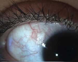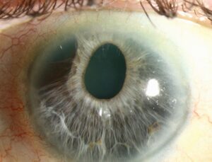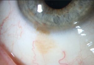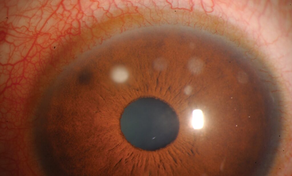
Subepithelial infiltrates are small gray-white corneal opacities that often follow adenoviral keratoconjunctivitis.
Patients call them viral spots, and most want to know when they will clear.
They usually fade, although the course can last many months.
Therefore, the visit centers on counseling and careful drug choices.
Why Subepithelial Infiltrates Form
The infiltrates typically appear one to two weeks after conjunctivitis begins.
They likely represent an immune reaction to viral antigen beneath the epithelium.
Inflammatory cells gather in the anterior stroma, just under Bowman layer.
As a result, active viral replication probably plays little role at this stage.
Typical Slit Lamp Appearance
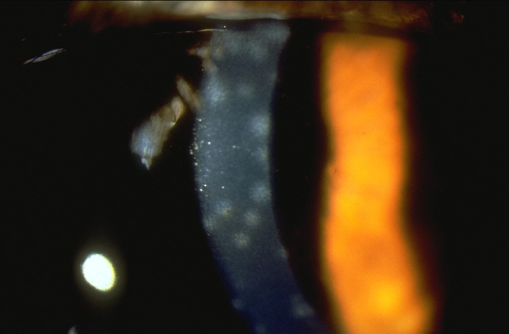
Multiple round, coin-shaped opacities sit mainly in the central cornea.
The overlying epithelium is usually intact, so fluorescein staining is minimal.
Retroillumination also outlines the dots clearly against the red reflex.
German authors call them nummuli because of this coin-like shape.
All-fit Slit-Lamp Adapter
Record and share exactly what you see at the slit lamp. One adapter fits any slit lamp or surgical microscope — and any smartphone.
From Choroida — the team behind this siteSymptoms and Glare in Subepithelial Infiltrates
Blurred vision dominates when lesions cross the visual axis.
Patients also describe glare, halos and photophobia, especially at night.
However, Snellen acuity may look nearly normal because the opacities mainly scatter light and reduce contrast.
Therefore, ask directly about night driving and screen work.
Irregular astigmatism over the lesions can further lower corrected acuity.
Do Subepithelial Infiltrates Clear?
In most patients, the infiltrates fade gradually over weeks to months.
A minority persists for a year or longer, with periodic recurrences.
Faint residual scars may remain, yet many cause no symptoms.
Then the real question becomes whether symptoms justify treatment.
Differential Diagnosis
History and lesion pattern help separate several lookalikes.
- Thygeson superficial punctate keratitis, with raised epithelial lesions and a quiet eye
- Herpes simplex stromal keratitis, often unilateral and recurrent
- Sterile infiltrates in contact lens wearers, usually peripheral
- Microbial keratitis, with pain and an epithelial defect
- Acanthamoeba keratitis in a lens wearer with severe pain (see Acanthamoeba ring infiltrate)
Observation First
Observation suits peripheral lesions and patients with good functional vision.
Preservative-free lubricants ease surface discomfort meanwhile.
In addition, sunglasses or tinted lenses reduce glare.
Clear reassurance prevents unnecessary visits and early drug escalation.
Most patients accept this plan once they understand the expected course.
Topical Steroids for Subepithelial Infiltrates
Topical corticosteroids suppress the infiltrates and often improve vision within days.
However, they do not shorten the underlying immune process.
Laibson and colleagues found that infiltrates often returned after steroid withdrawal.
Some authors also worry that early steroids may prolong viral shedding.
Therefore, reserve them for central lesions with real visual impact.
Steroid Rebound and Side Effects
Rebound causes most of the practical trouble.
Many patients become steroid dependent, relapsing whenever the dose falls too low.
Long courses also raise the risk of pressure elevation (see steroid-induced glaucoma).
Cataract and secondary infection add further concern over months.
As a result, measure pressure at every steroid visit.
Tapering Without Rebound
Choose a surface-acting agent such as loteprednol or fluorometholone when possible.
Then taper slowly over several weeks.
A gradual taper lowers rebound risk, although it cannot remove it.
If lesions recur, return to the lowest controlling dose.
Cyclosporine for Subepithelial Infiltrates
Topical cyclosporine offers a steroid-sparing route for persistent or recurrent lesions.
It inhibits T-cell activation, which fits the presumed immune mechanism.
Reinhard and colleagues reported benefit in a small pilot study.
Jeng and Holsclaw also described improvement in steroid-dependent eyes.
However, both reports were small and uncontrolled.
Cyclosporine Trade-Offs
Cyclosporine works slowly, so improvement may take weeks.
Burning on instillation is common and hurts adherence.
Higher concentrations often need compounding, which adds cost.
Therefore, many clinicians overlap it with a steroid, then withdraw the steroid gradually.
In return, it avoids pressure rise and lens changes.
Refractory Subepithelial Infiltrates
Topical tacrolimus appears in small case series for steroid-dependent disease.
Phototherapeutic keratectomy can address dense residual scars in selected eyes.
However, evidence for both remains limited.
Consequently, reserve them for visually significant, treatment-resistant cases.
Counseling and Follow-Up
Explain that the spots reflect immunity, with no active infection.
Describe the timeline in honest, flexible terms.
Warn steroid users about rebound before the first drop, because surprises damage trust.
Also record acuity, glare and slit lamp photos at each review.
Finally, ask patients to report pain or redness promptly.
Pitfalls to Avoid
- Starting steroids for peripheral lesions that barely affect vision
- Stopping steroids abruptly and triggering rebound
- Forgetting pressure checks during long courses
- Missing herpes simplex keratitis before prescribing steroids
- Promising a fixed clearance date
For the latest studies, see the PubMed results for subepithelial infiltrates.


Document what you see
Two smartphone imaging tools built for everyday clinic use — one for the slit lamp, one for the fundus.
From Choroida — the team behind this siteReferences
- Laibson PR, Dhiri S, Oconer J, Ortolan G. Corneal infiltrates in epidemic keratoconjunctivitis: response to double-blind corticosteroid therapy. Arch Ophthalmol. 1970;84:36-40.
- Jeng BH, Holsclaw DS. Cyclosporine A 1% eye drops for the treatment of subepithelial infiltrates after adenoviral keratoconjunctivitis. Cornea. 2011;30:958-961.
- Reinhard T, Godehardt E, Pfahl HG, Sundmacher R. Topical cyclosporin A in nummuli after keratoconjunctivitis epidemica: a pilot study. Ophthalmologe. 2000;97:764-768.
- Jhanji V, Chan TC, Li EY, Agarwal K, Vajpayee RB. Adenoviral keratoconjunctivitis. Surv Ophthalmol. 2015;60:435-443.
- Salmon JF. Kanski’s Clinical Ophthalmology: A Systematic Approach. 9th ed. Elsevier. 2020.

Writes and reviews the clinical articles on Educate. Every post is checked against peer-reviewed sources before it goes live.