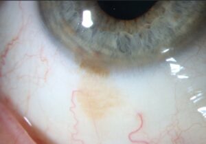
Pseudohypopyon is a layered deposit in the inferior anterior chamber composed of something other than inflammatory white cells.
It mimics hypopyon at first glance.
However, the causes include malignancy, retained lens material and injected drug crystals.
Therefore, every layered level in a quiet eye deserves a second look.
How Pseudohypopyon Differs From Hypopyon
A true hypopyon forms when leukocytes settle in an inflamed eye (see hypopyon in infection and uveitis).
The eye is usually painful, injected and full of flare.
By contrast, many pseudohypopyon cases arise in a white, comfortable eye.
As a result, a mismatch between the level and the inflammation is the first clue.
Why the Cause Matters
Mistaking a tumor for infection delays cancer treatment.
Also, an anterior chamber tap can seed retinoblastoma cells outside the eye.
Conversely, dismissing true endophthalmitis as a harmless deposit threatens the eye.
Therefore, each cause needs a deliberate workup.
All-fit Slit-Lamp Adapter
Record and share exactly what you see at the slit lamp. One adapter fits any slit lamp or surgical microscope — and any smartphone.
From Choroida — the team behind this siteRetinoblastoma Presenting With Pseudohypopyon
Retinoblastoma can shed tumor cells through the vitreous into the anterior chamber.
These cells settle as a white, chalky level, sometimes with small nodules.
The eye may look quiet, although secondary glaucoma can cause redness.
Therefore, a hypopyon in a young child without trauma or surgery should prompt exclusion of retinoblastoma.
Confirming Retinoblastoma
Examination under anesthesia often reveals the fundus mass or vitreous seeds (see retinoblastoma features).
B-scan ultrasound may show intralesional calcification, while MRI assesses the optic nerve.
However, clinicians generally avoid anterior chamber aspiration because of seeding risk.
Leukemia and Pseudohypopyon
Leukemic cells can infiltrate the iris and anterior chamber, sometimes as a sign of relapse.
The level may look gray-white and can mix with blood.
The iris may also look thickened or show new heterochromia.
In addition, cells blocking the trabecular meshwork may raise intraocular pressure.
Workup for Leukemic Infiltration
First, request a complete blood count with a peripheral smear.
Then, anterior chamber cytology can confirm malignant cells when doubt remains.
Also, involve the hematologist early, because ocular disease may change systemic treatment.
Lens Material After Cataract Surgery
Retained cortex or nuclear fragments can break down and layer in the anterior chamber.
The material looks fluffy, white and irregular.
It may also trigger inflammation or raised pressure (see lens-induced uveitis).
Gonioscopy often reveals the source fragment hiding in the inferior angle.
Separating Lens Material From Endophthalmitis
Both appear in the early postoperative period.
However, endophthalmitis usually brings worsening pain, lid swelling and falling vision.
Lens material tends to show discrete particles and a stable course.
When doubt persists, manage the eye as infected, because delay costs vision.
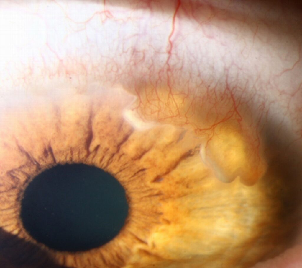
Triamcinolone Pseudohypopyon
Intravitreal triamcinolone crystals can migrate forward into the anterior chamber.
Migration seems more likely when the posterior capsule or zonules are not intact.
The level is white, crystalline and refractile, and it may shift with head position.
Also, the eye is usually quiet and comfortable.
Managing Triamcinolone Pseudohypopyon
Most deposits clear spontaneously over days to weeks.
Therefore, observation with close follow-up is usually enough.
Monitor intraocular pressure, because steroids raise it in susceptible eyes.
However, rising pain or vitreous haze needs urgent reassessment for infection.
Best Disease and Pseudohypopyon
In Best vitelliform macular dystrophy, the pseudohypopyon sits under the retina.
Yellow vitelliform material settles inferiorly within the macular lesion, creating a horizontal level.
OCT shows hyperreflective material layered in the lower subretinal space.
Vision is often better than the fundus appearance suggests.
In addition, electrooculography typically shows a reduced Arden ratio, even in carriers with normal fundi.
Other Causes of Pseudohypopyon
- Ghost cells after vitreous hemorrhage, which form a khaki layer
- Emulsified silicone oil, which floats upward as an inverse level
- Vitreoretinal lymphoma cells in older adults with steroid-resistant uveitis
- Pigment or tumor cells from necrotic uveal melanoma
A Practical Approach to Pseudohypopyon
- Note the color, texture and mobility of the level
- Compare the level with pain, injection and flare
- Ask about recent surgery, injections and cancer history
- Examine the fundus, or use B-scan when the view is poor
- Plan cytology only after excluding retinoblastoma
Counseling Patients With Pseudohypopyon
Explain that the deposit is a sign and its cause guides treatment.
For drug crystals, reassure the patient but list warning symptoms clearly.
For suspected malignancy, explain why referral is urgent.
Also, schedule review promptly, since several causes can change within days.
Pitfalls to Avoid
- Do not tap a child’s anterior chamber before excluding retinoblastoma
- Never assume a postinjection level is sterile without follow-up
- Remember that the Best disease level sits beneath the retina
For the latest studies, see the PubMed results for pseudohypopyon.


Document what you see
Two smartphone imaging tools built for everyday clinic use — one for the slit lamp, one for the fundus.
From Choroida — the team behind this siteReferences
- Shields CL, Shields JA. Diagnosis and management of retinoblastoma. Cancer Control. 2004;11:317-327.
- Kincaid MC, Green WR. Ocular and orbital involvement in leukemia. Surv Ophthalmol. 1983;27:211-232.
- Moshfeghi DM, Kaiser PK, Scott IU, et al. Acute endophthalmitis following intravitreal triamcinolone acetonide injection. Am J Ophthalmol. 2003;136:791-796.
- Roth DB, Chieh J, Spirn MJ, et al. Noninfectious endophthalmitis associated with intravitreal triamcinolone injection. Arch Ophthalmol. 2003;121:1279-1282.
- Boon CJF, Klevering BJ, Leroy BP, et al. The spectrum of ocular phenotypes caused by mutations in the BEST1 gene. Prog Retin Eye Res. 2009;28:187-205.
- Salmon JF. Kanski’s Clinical Ophthalmology: A Systematic Approach. 9th ed. Elsevier; 2020.

Writes and reviews the clinical articles on Educate. Every post is checked against peer-reviewed sources before it goes live.