Corneal endotheliitis is inflammation centered on the corneal endothelium, with stromal edema overlying keratic precipitates.
Herpes simplex virus is the classic cause, although cytomegalovirus and varicella zoster virus also produce it.
The epithelium usually stays intact, and the anterior chamber reaction is often mild.
As a result, the edema looks out of proportion to the inflammation.
What Defines Corneal Endotheliitis
The endothelial cell layer is the primary target.
Inflamed cells lose pump function, so fluid enters the stroma.
Therefore, the edema closely follows the inflamed area.
Keratic precipitates sit directly beneath the swollen zone, while stromal infiltrate and vessels are absent.
Three Clinical Forms
Holland and Schwartz described three patterns of herpetic endotheliitis.
- Disciform: a round zone of central or paracentral edema
- Diffuse: scattered precipitates with edema across the cornea
- Linear: a line of precipitates advancing from the limbus
All-fit Slit-Lamp Adapter
Record and share exactly what you see at the slit lamp. One adapter fits any slit lamp or surgical microscope — and any smartphone.
From Choroida — the team behind this siteDisciform Corneal Endotheliitis
Disciform disease is the most common form.
A sharp disc of stromal edema develops, often with epithelial microcysts.
Then, retroillumination reveals precipitates clustered beneath the disc.
Mild iritis may accompany it, and prognosis with treatment is generally good.
Diffuse Corneal Endotheliitis
In diffuse disease, precipitates scatter across the entire endothelium and edema involves the whole cornea.
As a result, vision usually drops more than in disciform disease.
Because the edema lacks a clear edge, clinicians may confuse it with dystrophic or surgical causes.
Linear Corneal Endotheliitis
Linear endotheliitis is the most aggressive form.
A line of precipitates starts at the limbus and moves toward the center.
Meanwhile, edema develops between the line and the limbus.
The picture resembles a Khodadoust rejection line, yet it occurs in ungrafted eyes.
Consequently, untreated disease can progress to endothelial decompensation.
Reading the KP Pattern Under the Edema
First, use a narrow slit beam and retroillumination.
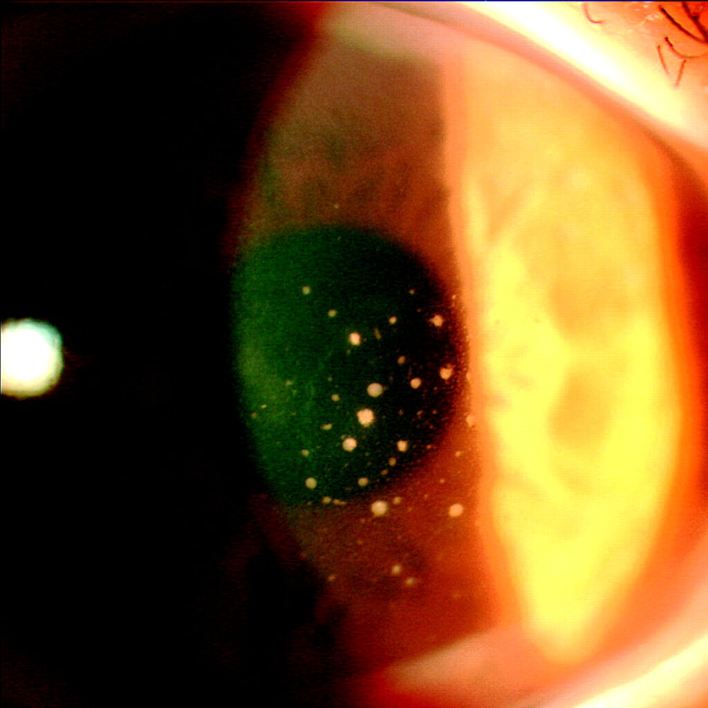
Fine or medium precipitates confined to the edematous zone suggest herpetic disease.
In addition, coin-shaped lesions (small precipitates arranged in a ring) strongly suggest cytomegalovirus.
For a broader guide, see keratic precipitate patterns.
HSV Corneal Endotheliitis
HSV disease is usually unilateral.
For example, many patients recall earlier dendritic ulcers or recurrent red eye.
Corneal sensation is also often reduced.
In addition, pressure may rise from trabeculitis.
However, a negative history does not exclude the diagnosis.
CMV Corneal Endotheliitis
CMV mostly affects immunocompetent middle-aged men, with many reports from Asia.
In particular, coin-shaped and linear precipitates are typical.
Also, pressure elevation is common and may be marked.
Recurrences are frequent, so endothelial cell loss accumulates.
CMV also appears after keratoplasty, where it mimics rejection (see viral anterior uveitis).
Confirming the Virus
Because clinical patterns overlap, aqueous humor PCR helps in atypical, recurrent or post-graft cases.
Koizumi and colleagues used this method to link CMV with endotheliitis.
Also, confocal microscopy can reveal enlarged owl’s eye cells in CMV disease.
Differential Diagnosis
- Corneal graft rejection with an endothelial rejection line
- Fuchs endothelial dystrophy with guttae
- Posner-Schlossman syndrome, which may itself be CMV related
- Herpes zoster endotheliitis with sectoral iris atrophy
Treating HSV Corneal Endotheliitis
Topical corticosteroids suppress the immune response that damages the endothelium.
The Herpetic Eye Disease Study showed that steroids hastened resolution of stromal keratitis.
However, steroids alone can reactivate epithelial disease.
Therefore, clinicians add antiviral cover, usually oral acyclovir or valacyclovir.
Linear disease often needs more frequent dosing and closer review.
Treating CMV Corneal Endotheliitis
Acyclovir has little activity against CMV, so clinicians choose ganciclovir-based therapy.
For example, options include topical ganciclovir gel, compounded drops or oral valganciclovir.
Then, a low-dose steroid controls inflammation.
Systemic valganciclovir can suppress the bone marrow, so blood counts need monitoring.
How Long Treatment Lasts in Corneal Endotheliitis
HSV disease usually needs weeks to months of tapering steroid.
Abrupt withdrawal can trigger rebound edema.
In addition, long-term oral acyclovir reduced recurrent stromal disease in the HEDS prevention trial.
CMV disease often needs many months of antiviral therapy.
Even then, recurrence after stopping is common, so some patients require maintenance.
Follow-Up and Outcomes
Review patients frequently during the first weeks.
Measure pressure at each visit, because both inflammation and steroids raise it.
Also, track central corneal thickness to document resolution.
Finally, persistent edema may need endothelial keratoplasty once the eye is quiet.
Counseling the Patient
Explain that the virus stays dormant and may return.
Patients should report blur, redness or halos early.
Long antiviral courses lower the risk, although they cannot eliminate recurrence.
Pitfalls to Avoid
- Do not label edema as graft rejection without first considering CMV
- Never stop steroids abruptly in herpetic disease
- Avoid steroids without antiviral cover in HSV cases
- Do not ignore pressure, because trabeculitis is common
- Remember that acyclovir does not treat CMV


Document what you see
Two smartphone imaging tools built for everyday clinic use — one for the slit lamp, one for the fundus.
From Choroida — the team behind this siteReferences
- Holland EJ, Schwartz GS. Classification of herpes simplex virus keratitis. Cornea. 1999;18:144-154.
- Wilhelmus KR, Gee L, Hauck WW, et al. Herpetic Eye Disease Study. A controlled trial of topical corticosteroids for herpes simplex stromal keratitis. Ophthalmology. 1994;101:1883-1896.
- Herpetic Eye Disease Study Group. Acyclovir for the prevention of recurrent herpes simplex virus eye disease. N Engl J Med. 1998;339:300-306.
- Koizumi N, Yamasaki K, Kawasaki S, et al. Cytomegalovirus in aqueous humor from an eye with corneal endotheliitis. Am J Ophthalmol. 2006;141:564-565.
- Koizumi N, Inatomi T, Suzuki T, et al. Clinical features and management of cytomegalovirus corneal endotheliitis: analysis of 106 cases from the Japan corneal endotheliitis study. Br J Ophthalmol. 2015;99:54-58.

Writes and reviews the clinical articles on Educate. Every post is checked against peer-reviewed sources before it goes live.