Corneal blood staining is the deposition of hemoglobin breakdown products in the corneal stroma after hyphema.
It usually follows a large or total hyphema that stays in the anterior chamber for days.
Raised intraocular pressure and a damaged endothelium drive most cases.
Because the opacity can persist for months, it matters most in young children.
What Corneal Blood Staining Is
Red cells in a hyphema release hemoglobin as they break down.
When the endothelial barrier fails, hemoglobin passes into the posterior stroma.
Keratocytes then take up the pigment and convert part of it to hemosiderin.
Histology also shows hemoglobin particles between collagen lamellae, which explains the brown color.
Risk Factors for Corneal Blood Staining
- Total or near-total (“eight-ball”) hyphema
- Intraocular pressure that stays elevated for several days
- Endothelial damage from the injury, prior surgery or dystrophy
- Rebleeding, which prolongs blood contact with the cornea
All-fit Slit-Lamp Adapter
Record and share exactly what you see at the slit lamp. One adapter fits any slit lamp or surgical microscope — and any smartphone.
From Choroida — the team behind this siteWhy Pressure and the Endothelium Matter
High pressure pushes hemoglobin across the endothelium and stresses its pump function.
A healthy endothelium, however, often resists staining even beside a large clot.
In contrast, a compromised endothelium may stain at near normal pressure.
Therefore, ask about previous intraocular surgery and Fuchs dystrophy (see corneal endothelial decompensation).
Early Slit-Lamp Signs of Corneal Blood Staining
The earliest change appears in the posterior stroma.
Examiners see fine yellowish granules and a loss of the normal fibrillar stromal detail.
The posterior stromal boundary then looks blurred on a narrow slit beam.
In addition, the cornea over the clot may take on a straw yellow tint.
These signs are subtle, so examine the patient daily while a total hyphema persists.
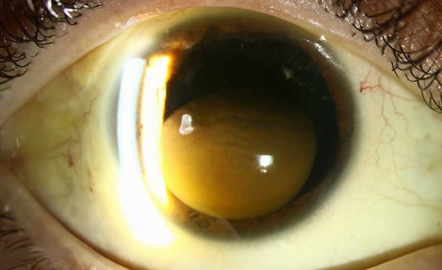
Established Corneal Blood Staining
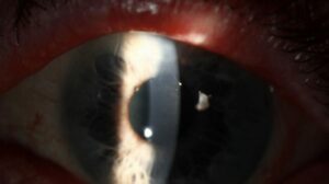
Later the stroma turns reddish brown, rust colored or greenish.
The opacity is often disc shaped and densest centrally.
A clearer rim commonly remains near the limbus.
As a result, the iris, lens and fundus may be hidden from view.
Differential Diagnosis
Blood resting on the endothelium can mimic early staining.
However, it shifts and clears as the hyphema resolves.
Other brown corneal changes include siderosis from a retained iron foreign body.
Also, keep the term separate from dye staining (see corneal staining patterns).
When to Wash Out the Hyphema
Early staining is an accepted indication for anterior chamber washout.
Classic guidance also favors surgery when a total hyphema persists with pressure around 25 mm Hg for about five days.
Washout removes the source of hemoglobin, but it cannot remove pigment already in the stroma.
Therefore, good timing depends on spotting the earliest signs.
Medical Management
Medical treatment aims to lower pressure and limit rebleeding.
Options include topical beta blockers, alpha agonists and topical steroids, with cycloplegia for comfort.
In sickle cell disease, clinicians usually avoid carbonic anhydrase inhibitors because they may promote sickling.
These measures reduce risk, while they do not dissolve established staining.
Clearing Pattern of Corneal Blood Staining
Staining resolves slowly, often over months and sometimes over two years or longer.
Clearing typically begins at the periphery and moves toward the center.
The visual axis is therefore the last area to clear.
The reason is uncertain, although closeness to the limbus may help pigment removal.
Amblyopia Risk in Children
In young children, a central opacity can cause deprivation amblyopia.
Because clearing takes months, the visual system may suffer during its sensitive period.
Therefore, involve a pediatric ophthalmologist early.
Occlusion of the fellow eye can then start once some central clarity returns.
Role of Keratoplasty
Most adults do well with observation, because the cornea usually clears on its own.
However, surgeons may consider keratoplasty when a dense central opacity threatens vision in an amblyogenic child.
Grafting carries rejection and failure risks, especially in a traumatized eye.
The decision then balances amblyopia against surgical risk.
Counseling the Patient
Explain that the brown color comes from blood pigment and does not mean infection.
Tell patients that clearing starts at the edges and may take a long time.
Also, explain that final vision depends on other injuries, such as angle recession or optic nerve damage.
Follow-Up in Corneal Blood Staining
Check pressure, corneal clarity and endothelial status at each visit.
Next, perform gonioscopy once the eye is quiet, because angle recession raises later glaucoma risk.
Finally, record the size of the clear zone to track progress.
Pitfalls to Avoid
- Do not wait for obvious brown color before acting
- Never assume normal pressure excludes staining when the endothelium is weak
- Do not overlook amblyopia in a child with a central opacity
For the latest studies, see the PubMed results for corneal blood staining.


Document what you see
Two smartphone imaging tools built for everyday clinic use — one for the slit lamp, one for the fundus.
From Choroida — the team behind this siteReferences
- Walton W, Von Hagen S, Grigorian R, Zarbin M. Management of traumatic hyphema. Surv Ophthalmol. 2002;47:297-334.
- Brodrick JD. Corneal blood staining after hyphaema. Br J Ophthalmol. 1972;56:589-593.
- McDonnell PJ, Green WR, Stevens RE, et al. Blood staining of the cornea: light microscopic and ultrastructural features. Ophthalmology. 1985;92:1668-1674.
- Read J, Goldberg MF. Comparison of medical treatment for traumatic hyphema. Trans Am Acad Ophthalmol Otolaryngol. 1974;78:799-815.
- Bowling B. Kanski’s Clinical Ophthalmology: A Systematic Approach. 8th ed. Elsevier; 2016.
- American Academy of Ophthalmology. Basic and Clinical Science Course, Section 8: External Disease and Cornea. San Francisco: AAO.

Writes and reviews the clinical articles on Educate. Every post is checked against peer-reviewed sources before it goes live.