Seidel test detects aqueous humor escaping through a full-thickness defect in the cornea, sclera or conjunctiva.
It uses concentrated fluorescein and cobalt blue light at the slit lamp.
The result often decides whether a wound needs urgent closure.
What the Seidel Test Shows
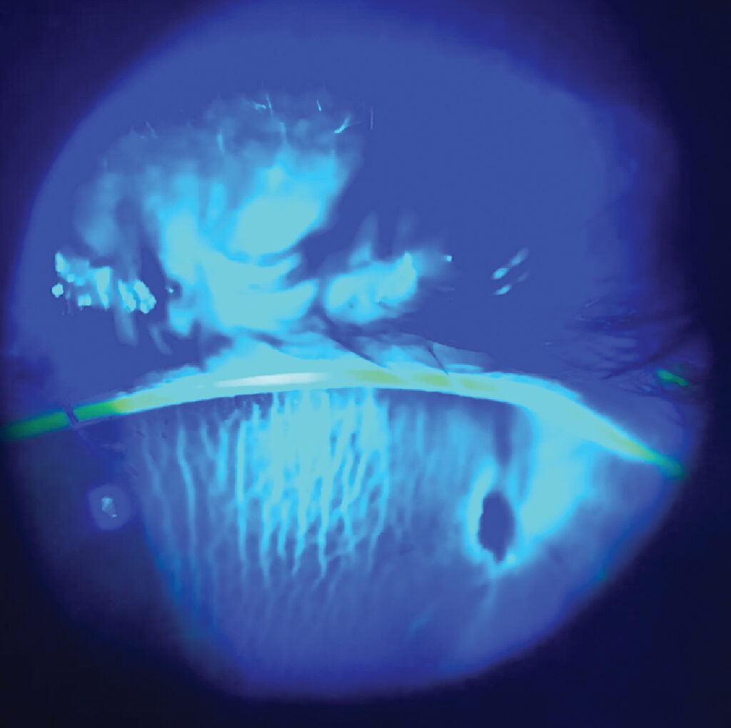
Concentrated fluorescein looks dark orange because its fluorescence is quenched.
However, when aqueous dilutes the dye, it glows bright green.
Therefore, a leak appears as a green stream flowing through the orange film.
Clinicians often call this stream a waterfall.
Equipment for the Seidel Test
- Sterile single-use fluorescein strips
- Sterile saline to moisten the strip
- Slit lamp with a cobalt blue filter
- Yellow barrier filter, if available, for better contrast
- Preservative-free anesthetic when the eye is painful
All-fit Slit-Lamp Adapter
Record and share exactly what you see at the slit lamp. One adapter fits any slit lamp or surgical microscope — and any smartphone.
From Choroida — the team behind this siteStep-by-Step Seidel Test Technique
- First, inspect the suspect area in white light for prolapsed tissue or a flat chamber.
- Next, wet the strip with one drop of saline.
- Then paint the strip gently over the site without pressing on the globe.
- Switch to cobalt blue light with a broad beam.
- Finally, watch the site steadily, because slow leaks may take a minute to appear.
Reading a Positive Seidel Test
A positive test shows a green rivulet streaming away from the site.
The stream usually runs downward with gravity.
Note whether the flow is brisk and continuous or slow and intermittent.
Also check chamber depth and intraocular pressure, because a brisk leak often lowers both.
Reading a Negative Result
A negative test shows an even orange film with no green stream.
However, a negative result does not prove that the wound is sealed.
Iris, vitreous or fibrin may plug the defect for a while.
In addition, very low intraocular pressure can stop the flow completely.
Pressure-Induced Leaks
Some wounds leak only when intraocular pressure rises.
In these eyes, gentle pressure through the lower lid may reveal a stream.
Record this as an induced positive result.
Never press on an obvious open globe, because pressure can expel intraocular contents.
Seidel Test After Corneal Laceration
In trauma, the test helps separate partial-thickness from full-thickness lacerations.
A positive result usually points toward primary closure (see corneal laceration repair).
However, a large obvious rupture needs no dye, because the diagnosis is already clear.
Small self-sealing wounds with a negative test may suit a bandage contact lens and close review.
Post-Operative Wound Leaks
After cataract surgery, a leaking incision can cause a shallow chamber and low pressure.
It also raises the risk of endophthalmitis, because tear film may enter the eye.
Therefore, test the main and side-port incisions in any eye with early hypotony (see clear corneal incision construction).
Small leaks often seal with a bandage lens, while persistent ones need a suture.
Seidel Test on a Filtering Bleb
After trabeculectomy, thin avascular blebs are a common source of leaks.
Paint the whole bleb surface, because pinpoint leaks hide easily.
Also separate a focal leak from diffuse oozing across a thin cystic bleb.
A leak with a shallow chamber or blebitis needs prompt glaucoma review.
Perforated Corneal Ulcers
A deep stromal ulcer can perforate with little warning.
The test confirms perforation, especially when the chamber shallows or iris moves toward the ulcer.
Small perforations often respond to tissue adhesive and a bandage lens (see corneal perforation management).
Larger defects usually need a patch graft or keratoplasty.
Seidel Test vs Corneal Staining
An epithelial defect takes up fluorescein and stays green in place.
A leak, however, shows green fluid moving away from its source.
Therefore, watch for flow before calling the test positive.
How the Result Changes Management
A brisk leak with a flat chamber usually needs urgent surgical closure.
A slow leak with a formed chamber may respond to conservative care first.
Options include a bandage contact lens, aqueous suppressants and frequent review.
Then repeat the test at each visit until the wound stays dry.
Document the site, flow speed and whether pressure was needed.
Counseling the Patient
Explain that the dye is safe and may tint tears yellow briefly.
Then describe the leak in plain words: fluid is escaping from inside the eye.
Advise the patient not to rub the eye and to wear a shield if supplied.
Finally, ask them to return urgently with pain, worse vision or discharge.
Pitfalls to Avoid
- Do not call epithelial staining a leak
- Do not trust one negative test in a hypotonous eye
- Avoid multidose fluorescein bottles near open wounds
- Do not soak the strip, because dilute dye removes the orange background
- Avoid reading too quickly, because intermittent leaks appear late


Document what you see
Two smartphone imaging tools built for everyday clinic use — one for the slit lamp, one for the fundus.
From Choroida — the team behind this siteReferences
- Romanchuk KG. Seidel’s test using 10% fluorescein. Can J Ophthalmol. 1979;14:253-256.
- Kuhn F, Morris R, Witherspoon CD, et al. A standardized classification of ocular trauma. Ophthalmology. 1996;103:240-243.
- Bowling B. Kanski’s Clinical Ophthalmology: A Systematic Approach. 8th ed. Elsevier; 2016.
- American Academy of Ophthalmology. Basic and Clinical Science Course, Section 8: External Disease and Cornea. San Francisco: AAO; 2023.
- Mannis MJ, Holland EJ, eds. Cornea: Fundamentals, Diagnosis and Management. 4th ed. Elsevier; 2017.

Writes and reviews the clinical articles on Educate. Every post is checked against peer-reviewed sources before it goes live.