An RPE tear is a rupture of the retinal pigment epithelium that can occur after anti-VEGF injection.
It happens most often under a large vascularized pigment epithelial detachment.
Therefore, it is a known risk in eyes with neovascular age-related macular degeneration.
What an RPE Tear Is
The pigment epithelium splits at the edge of a detachment.
The free edge retracts and rolls up, leaving bare Bruch’s membrane.
Also, the tear can involve the fovea.
As a result, vision may fall sharply.
Mechanism of an RPE Tear
Gass first described tears after laser treatment in 1984.
Fibrovascular tissue under the epithelium contracts after anti-VEGF drugs reduce the vascular component.
Also, the tissue shrinks and pulls the epithelium taut.
Therefore, the stress exceeds the strength of the layer and the epithelium tears.
Fundus Explorer Pro
Photograph the retinal findings described here with the phone already in your pocket — 22 D optics and built-in illumination in one handheld unit.
From Choroida — the team behind this siteIncidence
Studies report tears in a small share of eyes treated with anti-VEGF drugs.
The rate rises sharply in eyes with large vascularized detachments.
Also, a tear after a single injection is possible.
As a result, the risk per eye depends on baseline detachment size.
Risk Factors for an RPE Tear
A high pigment epithelial detachment, often above 550 micrometers, is the strongest predictor.
Large basal diameter and fibrovascular type add to it.
Also, Chan and colleagues and Doguizi and Ozdek described the link with detachment size.
Therefore, measure the height and width of the detachment before treatment.
Timing
Most tears occur within the first three injections.
Many appear within a few weeks of the first or second dose.
Also, late tears are rare.
In addition, a sudden fall in vision after an injection should prompt a scan.
Clinical Appearance
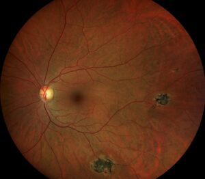
The fundus shows a crescent of bare choroid next to a dark, rolled edge of pigment.
Subretinal hemorrhage may accompany it.
Also, fluorescein angiography shows early hyperfluorescence of the bare area.
As a result, the diagnosis is usually clear on examination.
Imaging an RPE Tear
OCT shows a retracted, folded epithelium and an exposed Bruch’s membrane.
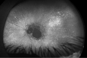
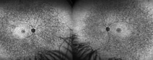
Fundus autofluorescence shows a dark area of absent epithelium with a bright rolled edge.
Also, the detachment shrinks on the scan.
Therefore, multimodal imaging confirms the tear.
Visual Outcome
Prognosis depends on foveal involvement.
Tears that spare the fovea often keep good vision.
Also, tears that involve the fovea cause lasting loss.
Therefore, the location of the tear sets the outcome.
Continuing Anti-VEGF Therapy
Most experts continue treatment after a tear because the neovascular disease remains active.
Studies show that vision stabilizes in many eyes with continued injections.
Also, the interval may be adjusted to control exudation.
As a result, a tear is not a contraindication.
Preventing an RPE Tear
There is no sure prevention.
Some clinicians warn patients about the risk and use a gentle loading regimen in high detachments.
Also, Sarraf and colleagues compared low and high doses and found no clear difference in tear rates.
In addition, careful follow-up allows early detection.
Counseling Patients
Explain before the first injection that tears can occur and may cause a visible drop in vision.
Ask them to report sudden loss within days of an injection.
Also, reassure them that treatment may continue.
Therefore, informed consent includes this risk.
Fellow Eye
Check the fellow eye for large detachments.
A tear in one eye predicts a higher risk in the other.
Also, monitor with OCT at each visit.
As a result, early warning signs may be caught.
Differential Diagnosis
A subretinal hemorrhage can mimic a tear but lacks the bare area.
Geographic atrophy shows a bare area without a rolled edge (see geographic atrophy treatment).
Also, a macular hole shows a retinal defect on OCT.
Therefore, OCT and autofluorescence separate the entities.
Surgical Options
Surgery has a limited role.
Vitrectomy may help when hemorrhage is dense.
Also, no surgery repairs the tear itself.
In addition, results of submacular surgery have been poor.
Pitfalls to Avoid
- Do not stop treatment after a tear without discussing the risk of exudation
- Never ignore the height of the detachment before the first injection
- Do not forget to examine the fellow eye
- Never skip autofluorescence in a suspected tear
Natural Course Without Treatment
Without treatment, many eyes with a tear continue to leak and lose vision.
The neovascular lesion usually stays active.
Also, the bare area can enlarge slowly.
Therefore, treatment decisions should weigh both risks.
For the latest studies, see the PubMed results for RPE tear.


Document what you see
Two smartphone imaging tools built for everyday clinic use — one for the slit lamp, one for the fundus.
From Choroida — the team behind this siteReferences
- Gass JDM. Retinal pigment epithelial rip during krypton red laser photocoagulation. Am J Ophthalmol. 1984;98:700-706.
- Chan CK, Meyer CH, Gross JG, et al. Retinal pigment epithelial tears after intravitreal bevacizumab injection for exudative age-related macular degeneration. Retina. 2007;27:541-551.
- Doguizi S, Ozdek S. Pigment epithelial tears associated with anti-VEGF therapy: incidence, long-term visual outcome, and relationship with pigment epithelial detachment in age-related macular degeneration. Retina. 2014;34:1156-1162.
- Sarraf D, Chan C, Rahimy E, Abraham P. Prospective evaluation of the incidence and risk factors for the development of RPE tears after high- and low-dose ranibizumab therapy. Retina. 2013;33:1551-1557.

Writes and reviews the clinical articles on Educate. Every post is checked against peer-reviewed sources before it goes live.