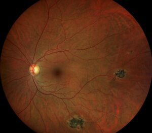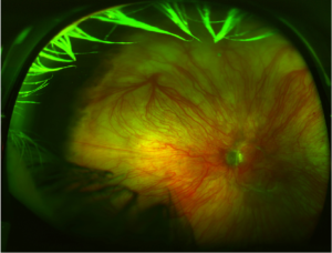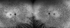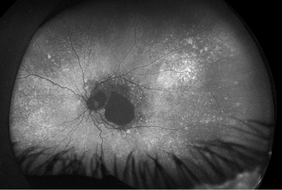
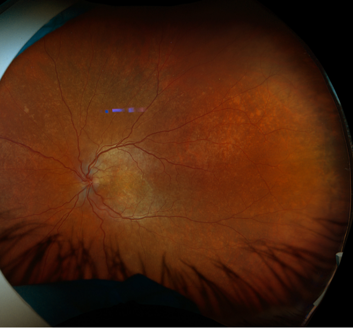
A salt-and-pepper fundus shows fine, evenly scattered dark and light spots across the retina.
The spots come from the retinal pigment epithelium, which alternates between clumped and depigmented patches.
Because several infections and dystrophies produce this look, the pattern needs a systematic workup.
What a Salt-and-Pepper Fundus Looks Like
The change is diffuse and usually most obvious in the posterior pole and the midperiphery.
The vessels and the optic disc often look normal.
Also, vision is frequently good, which surprises families and clinicians alike.
Fundus autofluorescence shows a mottled pattern that matches the pigment change.
Infectious Causes of a Salt-and-Pepper Fundus
- The best known cause is congenital rubella (see congenital rubella syndrome)
- Syphilis acquired before birth, which may also cause interstitial keratitis
- Cytomegalovirus and measles, in rarer cases
Givens and colleagues found that rubella retinopathy is usually benign and does not progress.
However, a subretinal neovascular membrane can appear years later.
Fundus Explorer Pro
Photograph the retinal findings described here with the phone already in your pocket — 22 D optics and built-in illumination in one handheld unit.
From Choroida — the team behind this siteHereditary Causes of a Salt-and-Pepper Fundus
Carriers of X-linked retinitis pigmentosa may show a similar mottled change.
Early retinitis pigmentosa and Cockayne syndrome can also produce it.
In these patients, the electroretinogram helps to separate a stable pigmentary change from a progressive dystrophy (see electroretinography).
Workup for a Salt-and-Pepper Fundus
First, take a maternal history of rubella, syphilis, and other infections in pregnancy.
Then order treponemal serology, because ocular syphilis mimics many conditions (see ocular syphilis).
Add electroretinography when night vision or the field is abnormal.
Finally, examine the parents, since carrier states may be visible.
Prognosis and Follow-Up of a Salt-and-Pepper Fundus
Most children with rubella retinopathy keep good central vision for life.
However, the other manifestations of congenital rubella matter more for the child, including cataract, glaucoma, deafness, and heart defects.
Therefore, ask about cardiac and hearing assessments, and refer when they are missing.
Review the patient yearly, and teach older children to report distortion or a sudden drop in vision.
Neovascular membranes are uncommon, but they respond to anti-VEGF injections when they occur.
Congenital Syphilis and a Salt-and-Pepper Fundus
Congenital syphilis can look identical to rubella retinopathy in the fundus.
Also, the child may have interstitial keratitis, deafness, and dental changes.
Confirm the diagnosis with treponemal and nontreponemal serology, and refer for neurosyphilis assessment.
Treat with intravenous penicillin under pediatric infectious disease care.
Because maternal infection is the source, arrange testing and treatment for the mother and her partner.
Salt-and-Pepper Fundus Versus Retinitis Pigmentosa
Rubella retinopathy is stationary, and the vessels and disc remain normal.
In contrast, retinitis pigmentosa progresses, narrows the arterioles, and produces bone spicules (see retinitis pigmentosa).
The electroretinogram is normal or only mildly reduced in rubella, whereas it declines steadily in retinitis pigmentosa.
Also, night vision is usually preserved in rubella.
Therefore, a normal electroretinogram with a mottled fundus favors a congenital infection or a carrier state.
Prevention of Congenital Rubella
Vaccination prevents the disease, and congenital rubella has become rare where coverage is high.
However, pockets of low coverage still produce cases.
Ask about maternal vaccination and travel history in every child with the pattern.
Also report suspected cases to public health authorities.
Finally, counsel women of childbearing age to check their immunity before pregnancy.
Practical Points for the Clinician
Examine both eyes with dilation, and look closely at the macula for any membrane or hemorrhage.
Also measure the refraction, since amblyopia and high myopia occur in some children.
Photograph the fundus, because the mottling is easy to miss on later review.
Then correlate the finding with the systemic history, and avoid labeling the child with a progressive disease without proof.
Communicating With Families
Explain that the fundus change is usually stable and rarely threatens sight.
Also describe the other organ systems that need follow-up.
Because parents often fear blindness, state clearly what the electroretinogram shows.
Finally, review warning signs such as distortion, a new blind spot, or a drop in vision.
Imaging and Electrophysiology
Autofluorescence shows a speckled pattern that matches the pigment change.
Optical coherence tomography usually shows a normal macular profile with mild outer retinal irregularity.
Also, full-field electroretinography is normal or slightly reduced in rubella.
Therefore, abnormal responses should prompt a search for a second diagnosis.
Order the test when the child can cooperate, or arrange it under sedation when needed.
Other Clues in the History
Ask about hearing screening results, because deafness accompanies rubella and syphilis.
Also ask about heart surgery in infancy, which suggests congenital rubella.
Review the birth record for low weight and small head size.
Then check developmental milestones, since neurologic involvement changes the plan.
For the latest studies, see the PubMed results for salt-and-pepper fundus.


Document what you see
Two smartphone imaging tools built for everyday clinic use — one for the slit lamp, one for the fundus.
From Choroida — the team behind this siteReferences
- Givens KT, Lee DA, Jones T, Ilstrup DM. Congenital rubella syndrome: ophthalmic manifestations and associated systemic disorders. Br J Ophthalmol. 1993;77:358-363.
- Tamesis RR, Foster CS. Ocular syphilis. Ophthalmology. 1990;97:1281-1287.
- Hartong DT, Berson EL, Dryja TP. Retinitis pigmentosa. Lancet. 2006;368:1795-1809.
- Wolff SM. The ocular manifestations of congenital rubella: a prospective study of 328 cases of congenital rubella. Trans Am Ophthalmol Soc. 1972;70:577-614.

Writes and reviews the clinical articles on Educate. Every post is checked against peer-reviewed sources before it goes live.